FIGURE SUMMARY
Title

Shigella serotypes associated with carriage in humans establish persistent infection in zebrafish

Authors
Torraca, V., Brokatzky, D., Miles, S.L., Chong, C.E., De Silva, P.M., Baker, S., Jenkins, C., Holt, K.E., Baker, K.S., Mostowy, S.
Source
Full text @ J. Infect. Dis.

Shigella establishes persistent infection in zebrafish. A, Experimental diagram of S. sonnei–zebrafish infection. S. sonnei infection undergoes 3 phases: an acute phase (ie, increasing bacterial load), a clearing phase (ie, a steady decrease of bacterial load), and a persistent phase (ie, bacterial load does not further decrease). Diagram created with BioRender.com. B and C, CFU count and survival analysis from zebrafish larvae infected with S. sonnei 53G. Larvae were treated with the antibiotic Nal or left untreated. S. sonnei establishes a persistent infection in zebrafish, even during treatment with a therapeutic antibiotic dose. From 96 hpi, the bacterial load in larvae remains constant over consecutive days. D, CFU count from zebrafish larvae treated with Nal or left untreated during the acute and persistent infection stages. During the acute infection stage (0–24 hpi), S. sonnei is sensitive to antibiotic treatment, while during the persistent infection stage (120–144 hpi), S. sonnei becomes insensitive to antibiotic treatment. Data were analyzed at 24 hours posttreatment. Data: mean ± SEM. Statistics: B, unpaired t test on log10-transformed data against the previous time point; C, log-rank Mantel-Cox test; D, 1-way analysis of variance with Sidak correction. ns, P ≥ .05. *P < .05. ***P < .001. CFU, colony-forming unit; hpi, hours postinfection; hpt, hours posttreatment; Nal, nalidixic acid; ns, nonsignificant.

Persistent Shigella represents a clonally expanded population. mCherry- and GFP-labeled S. sonnei 53G strains were coinjected at a 1:1 ratio. A, At 0 hpi, most larvae (approximately 90%) displayed an expected 1:1 ratio of the coinjected strains (ie, percentage difference between strains ≤20%). B, At 144 hpi, most larvae (approximately 80%) were infected by 1 of the 2 strains at a much higher frequency than the other strain (ie, percentage difference between strains >80%), indicating the establishment of clonality. Statistics: differences in distribution between data at 0 and 144 hpi were calculated by chi-square test. ****P < .0001. hpi, hours postinfection.

Shigella O-antigen is essential to establish persistent infection. A and B, Percentage of larvae carrying persistent infection at 144 hpi and average bacterial log10CFU for wild type S. sonnei 53G and several isogenic mutants. A T3SS mutant (ΔMxiD) can establish persistent infection at similar levels as wild type. However, the –pSS mutant (depleted of the virulence plasmid) and an O-antigen mutant (ΔO-Ag) are significantly reduced in their ability to establish persistent infections. C and D, Percentage of larvae carrying persistent infection at 144 hpi and average bacterial log10CFU for several S. sonnei isolates from lineage II and lineage III. Lineage II and III S. sonnei isolates are both capable of establishing persistent infections. Although belonging to different lineages, all S. sonnei isolates share an identical O-antigen. Data: mean ± SEM. Statistics: 1-way analysis of variance with Sidak correction on (A, C) percentage data or (B, D) log10-transformed data. ns, P ≥ .05. **P < .01. ***P < .001. ****P < .0001. CFU, colony-forming unit; hpi, hours postinfection; ns, nonsignificant; T3SS, type III secretion system; WT, wild type.

Phylogenetic distribution of persistent Shigella. Phylogenetic trees (codon trees) for (A) S. sonnei and (B) S. flexneri were constructed by using complete reference genomes of known serotypes (available in BV-BRC, https://www.bv-brc.org/), draft genome sequences of isolates associated with persistent carriage in humans [29], and the isolates used in this study (sequenced with nanopore technology). Red (labeled with diamond symbols ◆), strains associated with persistent carriage in humans; orange (labeled with star symbols ★), strains that established persistent infection in zebrafish; black (labeled with square symbols ■), strains that did not establish persistent infection in zebrafish; blue (unlabeled), other strains available in the BV-BRC database and utilized to construct the phylogenetic trees. For the S. sonnei tree, lineages II and III and sublineages 3.6 and 3.7 are highlighted. For the S. flexneri tree, the 3 main clusters encompassing the serotypes investigated in this study (2a, 3, and 5) are highlighted, and the serotype of each strain is reported in parentheses after the strain name. BV-BRC, Bacterial and Viral Bioinformatics Resource Centre.

Shigella O-antigen serotypes associated with MSM transmission enable persistent infection. A and B, Percentage of larvae carrying persistent infection at 144 hpi and average bacterial log10CFU for several S. flexneri isolates from serotypes 2a, 3a, and 5a. S. flexneri isolates of serotypes 2a and 3a are both capable of establishing persistent infection, to a much greater extent than S. flexneri of serotype 5a. C and D, Percentage of larvae carrying persistent infection at 144 hpi and average bacterial log10CFU for S. flexneri M90T (serotype 5a), S. sonnei 53G, and S. flexneri SRR12769770 (a 2020 clinical isolate of S. flexneri from serotype 5a). SRR12769770 does not show a significant difference in the establishment of persistent infection when compared with S. flexneri M90T. Data: mean ± SEM. Statistics: 1-way analysis of variance with Sidak correction on (A, C) percentage data or (B, D) log10-transformed data. ns, P ≥ .05. **P < .01. ***P < .001. ****P < .0001. CFU, colony-forming unit; hpi, hours postinfection; MSM, men who have sex with men; ns, nonsignificant.

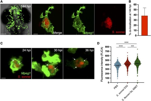
Shigella can establish persistent infection of macrophages in vivo. A, Head region and individual macrophage detail from a representative S. sonnei–infected zebrafish larva at 144 hpi. Tg(mpeg1::Gal4-FF)gl25/Tg(UAS::LIFEACT-GFP)mu271 larvae (Mpeg1, with macrophages in green) were injected at 3 dpf in the hindbrain ventricle with 1000 CFU of mCherry-labeled S. sonnei 53G (red). An infected macrophage harboring persistent bacteria is magnified. Scale bars: 100 μm, left; 10 μm, right and inset. B, At the persistent infection stage, 40% of bacterial fluorescence colocalizes with mpeg1+ macrophages. C, Individual Tg(mpeg1::Gal4-FF)gl25/Tg(UAS::LIFEACT-GFP)mu271 macrophage (Mpeg1, green) harboring mCherry–S. sonnei (red, indicated by arrows) followed for 12 hours (from 24 to 36 hpi). At 2 dpf, 1000 CFU of bacteria were delivered systemically in larvae. Scale bar: 10 μm. D, As compared with S. flexneri M90T, S. sonnei 53G induces a reduced level of caspase 1 activation in vivo in the zebrafish model. At 3 dpf, 10000 CFU of bacteria were delivered systemically in larvae. Data: mean ± SEM. Statistics: 1-way analysis of variance with Sidak correction. ***P < .001. ****P < 0.0001. CFU, colony-forming unit; dpf, days postfertilization; hpi, hours postinfection; PBS, phosphate-buffered saline.

Acknowledgments
This image is the copyrighted work of the attributed author or publisher, and ZFIN has permission only to display this image to its users. Additional permissions should be obtained from the applicable author or publisher of the image. Full text @ J. Infect. Dis.