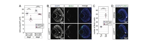
The retina of cct5𝑡𝑓121𝑏mutants is smaller and characterised by enhanced proliferation and apoptosis. (A) Quantification of the retina sizes revealed that the retinal area of cct5𝑡𝑓121𝑏 homozygotes was significantly smaller compared to siblings. The mean retinal area of cct5𝑡𝑓121𝑏 homozygotes was 17.8 ± 0.8 (×103 μm2) at three dpf and 23.8 ± 0.2 (×103 μm2) at six dpf compared to siblings that featured mean retinal areas of 24.7 ± 0.5 (×103 μm2) at three dpf and 28.4 ± 0.5 (×103 μm2) at six dpf; n = 10 per genotype and stage. (B) In contrast to three-dpf-old siblings, more BrdU-positive cells (green) were detected at the periphery of the retina within the ciliary marginal zone of cct5𝑡𝑓121𝑏 homozygotes. (C) Quantification of the number of BrdU-positive cells within individual retinas revealed that significantly more cells were proliferating within the ciliary marginal zone of cct5𝑡𝑓121𝑏 homozygotes at three dpf. Whereas cct5𝑡𝑓121𝑏 had 48 ± 3 BrdU-positive cells per retinal cross section, 18 ± 1 were proliferating in siblings (n = 5). (D) In contrast to the retina of three-dpf-old siblings, in which apoptotic cells were rarely labelled by the TUNEL assay (green), apoptosis was frequently observed throughout the entire retina of cct5𝑡𝑓121𝑏homozygotes. Data are mean ± SEM; ***P < 0.001 by Student’s t-test. Arrowheads indicate optic nerves (ON).
|
