Fig. 7
- ID
- ZDB-FIG-221114-13
- Publication
- Wang et al., 2021 - atg7-Based Autophagy Activation Reverses Doxorubicin-Induced Cardiotoxicity
- Other Figures
- All Figure Page
- Back to All Figure Page
|
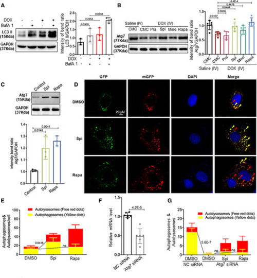
Spironolactone (Spi) and rapamycin (Rapa) activated autophagosome formation in an Atg7-dependent fashion. A, Representative Western blot and quantification of the relative amounts of LC3-II (microtubule-associated protein light chain 3II) in the hearts from mice injected with a single bolus of doxorubicin (DOX). Activated LC3-II and an increased response to bafilomycin A1 (BafA1; n=5/treatment) were observed. B, Representative Western blots and quantification of Atg7 from the hearts of mice administered the 4 drugs daily in the later phase. C, Representative Western blot and quantification of Atg7 from the H9C2 cardiac cell line. Spi and Rapa induced an increase in Atg7 protein levels. D, H9C2 cells were transiently transfected with mRFP-green fluorescence protein (GFP)-LC3 adenovirus and then treated with Spi and Rapa. Representative images of GFP-LC3 and mRFP-LC3 puncta are shown. E, Quantification of the yellow puncta (autophagosomes) and red puncta (autolysosomes) is shown in D. F, Quantitative real-time polymerase chain reaction was used to confirm that Atg7 transcripts were reduced by Atg7 siRNA. G, Quantitative analysis of the yellow puncta (autophagosomes) and red puncta (autolysosomes) showing that Atg7 siRNA ablates the induction of autophagosomes induced by Spi or Rapa. Kruskal-Wallis test was used followed by post hoc Tukey test in A and C; 1-way ANOVA followed by post hoc Tukey test in B, E, and G; Student test in F. CMC indicates carboxymethylcellulose; DMSO, dimethyl sulfoxide; GFP, green fluorescence protein; H9C2, rat cardiomyocytes; IV, intravenous; LC3, light chain 3; Mino, minoxidil; mGFP, monomeric green fluorescence protein; mRFP, monomeric red fluorescence protein; NC, negative control; Pra, pravastatin; and SiRNA, small interfering RNA. |

