
Fig. 7
- ID
- ZDB-FIG-221030-51
- Publication
- Zheng et al., 2021 - Molecular basis for bipartite recognition of histone H3 by the PZP domain of PHF14
- Other Figures
- All Figure Page
- Back to All Figure Page
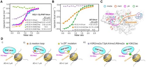
Modification crosstalk and hierarchical switch of H3-PHF14PZP engagement. (A,B) ITC fitting curves of H3(1–13)-PHF14PZP (A) and AF10PZP (B) titrated with H3(14–34) peptides with indicated modifications. N.D. = not detected. (C) A model illustrating modification switch features of H3(1–34)-PHF14PZP engagement. H3-Nter and H3-middle are colored magenta and orange, respectively. Indicated modifications are shown as color-coded spheres. Strictly sensitive, mildly sensitive, and tolerated modifications are denoted by red cross, half-tick, and tick signs, respectively. (D) A model highlighting hierarchical modification switch features for downregulation of the H3(1–34)-PHF14PZP complex formation. H3-Nter and H3-middle are colored magenta and orange, respectively. Hexagonal stars of different colors indicate protein mutations at insertion loop region, mutation at ‘α-ZP’ surface, histone modifications in H3-Nter, and histone modification in H3-middle region, respectively. |