
Fig. 4
- ID
- ZDB-FIG-221030-48
- Publication
- Zheng et al., 2021 - Molecular basis for bipartite recognition of histone H3 by the PZP domain of PHF14
- Other Figures
- All Figure Page
- Back to All Figure Page
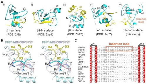
A novel recognition model for H3 peptide readout by PHF14PZP. (A) A summary of different recognition modes of PHD fingers. PHD fingers and their ligands are depicted by cyan and yellow ribbons, respectively. Zinc ions are shown as white spheres and the corresponding secondary structure are labelled. (B) Structural alignment of H3 (yellow)-PHF14 (pale cyan), H3 (light pink)-BHC80 (blue white) and H3 (cyan)-BPTF (white) complexes in wall-eye stereo view. H3 R2, K4, and R8 are depicted as sticks. (C) Sequence alignment of different PHD finger proteins: BHC80 (2PUY), PYGO (2VP7), ING2 (2G6Q), BPTF (2F6J), UHRF1 (3SOX), TAF (5WXG), MLL4PHD6 (6O7G), AIRE (2KFT), MOZ (5B78), BRPF1 (6U04), TRIM24 (3O37), AF10 (5DAH), ORC1b (5HH7). |