FIGURE

Fig. 6.

ID
ZDB-FIG-220317-13
Publication
Serrano et al., 2021 - Novel pre-clinical model for CDKL5 Deficiency Disorder
Other Figures
All Figure Page
Back to All Figure Page
Fig. 6.

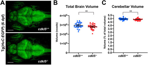
Brain imaging in cdkl5−/− fish. (A) Maximum intensity projections of confocal images from whole brains from Tg(HuC:EGFP); cdkl5+/+ and Tg(HuC:EGFP); cdkl5−/− fish at 6 dpf. Scale bars: 100 µm. (B,C) Quantification of total brain (B) and cerebellar (C) volume from cdkl5+/+ and cdkl5−/− fish. Data are mean±s.e.m. for three independent experiments (n=12, 12 and 11 for cdkl5+/+, and n=12, 12 and 12 for cdkl5−/− fish), **P<0.01 (two-tailed t-test). 1 voxel=8 µm3.

Expression Data
Gene:
Fish:
Anatomical Term:
Stage: Day 6

Expression Detail
Antibody Labeling
Phenotype Data
Fish:
Observed In:
Stage: Day 6

Phenotype Detail
Acknowledgments
This image is the copyrighted work of the attributed author or publisher, and ZFIN has permission only to display this image to its users. Additional permissions should be obtained from the applicable author or publisher of the image. Full text @ Dis. Model. Mech.