FIGURE SUMMARY
Title

Novel pre-clinical model for CDKL5 Deficiency Disorder

Authors
Serrano, R.J., Lee, C., Douek, A.M., Kaslin, J., Bryson-Richardson, R.J., Sztal, T.E.
Source
Full text @ Dis. Model. Mech.

cdkl5 expression and validation of the cdkl5sa21938 zebrafish mutant strain. (A) Schematic of the zebrafish Cdkl5 long (Cdkl5-L) and short (Cdkl5-S) protein isoforms showing the location of the nonsense (PTC) mutation and primers used for RT-PCR analyses. The long (3936 bp) and short isoforms (3479 bp) both contain exons 1-15 and exons 17-18 (grey), and differ by the inclusion of exon 16 (white) and the lengths of their 5′ untranslated region (UTR) (orange) and 3′ UTR (green) regions. (B) RT-PCR analyses of cdkl5-L and cdkl5-S transcripts from 1-6 dpf.  β-Act was amplified as a positive control. (C) cdkl5 mutants contain a C>A change in exon 11 of the gene (amino acid 528 out of 1039 and 1080 of the short and long isoforms, respectively), creating a premature termination codon (PTC), as seen in the sequencing chromatograms from cdkl5+/− fish. (D) qRT-PCR analysis demonstrated that cdkl5-L and total cdkl5 mRNA levels are significantly reduced in cdkl5−/− embryos compared to cdkl5+/+ siblings at 3 dpf. Data are mean±s.e.m. for three independent experiments (with 20 pooled fish per experiment), *P<0.05 (one-way ANOVA).

EXPRESSION / LABELING:
Gene:
Fish:
Anatomical Term:
Stage Range: Prim-5 to Day 6

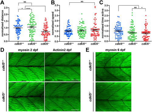
Motor function and muscle pathology in cdkl5−/− fish. (A-C) Quantification of normalized distance travelled (A), speed (B) and time active (C) of cdkl5+/+, cdkl5+/− and cdkl5−/− fish at 6 dpf. Data are mean±s.e.m. for three independent experiments (n=38, 21 and 15 for cdkl5+/+; n=17, 39 and 19 for cdkl5+/−; and n=27, 25 and 14 for cdkl5−/− fish per experiment). *P<0.05; ****P<0.0001; NS, not significant (one-way ANOVA). (D) Maximum intensity projection of a confocal image series of α-myosin and α-Actinin2 antibody staining of cdkl5+/+ and cdkl5−/− fish at 2 dpf. (E) Maximum intensity projections of confocal images of α-myosin antibody staining of cdkl5+/+ and cdkl5−/− fish at 6 dpf. Scale bars: 100 µm (D); 10 µm (E).

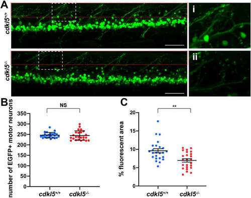
Assessment of neuronal pathology in cdkl5−/− fish. (A) Maximum intensity projection confocal images of EGFP-labelled motor neurons from Tg(islet1:EGFP); cdkl5+/+ (white dashed box zoomed in i) and Tg(islet1:EGFP); cdkl5−/− (white dashed box zoomed in ii) along the spinal region at 6 dpf. Red dashed boxes indicate the regions quantified in C. Scale bars: 50 µm. (B) Quantification of the number of EGFP+ motor neurons at 6 dpf. Data are mean±95% c.i. for three independent experiments [n=8, 8 and 8 for Tg(islet1:EGFP); cdkl5+/+, and n=8, 8 and 8 for Tg(islet1:EGFP); cdkl5−/− fish]. (C) Quantification of the mean percentage area of fluorescence from Tg(islet1:EGFP); cdkl5+/+ and Tg(islet1:EGFP); cdkl5−/− (within the red dashed box region in A) at 6 dpf. Data are mean±s.e.m. for three independent experiments [n=8, 8 and 8 for Tg(islet1:EGFP); cdkl5+/+, and n=8, 8 and 8 for Tg(islet1: EGFP); cdkl5−/− fish]. **P<0.01; NS, not significant (two-tailed t-test).

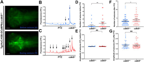
Analysis of seizures in cdkl5−/− fish. (A) Images of the brain region in Tg(HuC:H2B-GCaMP6s); cdkl5+/+ fish before (indicated in B by the blue arrowhead) and during a seizure (indicated in B by the blue arrow) at 6 dpf. Scale bars: 100 µm. (B,C) Representative time trace images of fluorescence (F/F0,) in control conditions and after the addition of 20 mM PTZ (at the time indicated) for Tg(HuC:H2B-GCaMP6s);cdkl5+/+ (B) and Tg(HuC:H2B-GCaMP6s);cdkl5−/− (C) fish. The fluorescence data are integrated over the whole embryonic brain. Seizures (arrows) are represented by peaks in fluorescence that extend above the baseline level (represented by the dotted line). (D,E) Quantification of the number (D) and amplitude (E) of seizures observed before PTZ treatment in Tg(HuC:H2B-GCaMP6s);cdkl5+/+ and Tg(HuC:H2B-GCaMP6s);cdkl5−/− fish. (F,G) Quantification of the number (F) and amplitude (G) of seizures observed after PTZ treatment in Tg(HuC:H2B-GCaMP6s);cdkl5+/+ and Tg(HuC:H2B-GCaMP6s);cdkl5−/− fish. Data are mean±95% c.i. for three independent experiments [n=9, 12 and 18 for Tg(HuC:H2B-GCaMP6s);cdkl5+/+, and n=19, 13 and 16 for Tg(HuC:H2B-GCaMP6s);cdkl5−/− fish]. *P<0.05; **P<0.01; NS, not significant (two-tailed t-test).

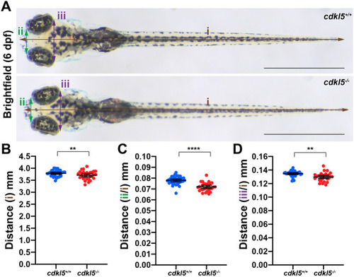
Analysis of body length and head size in cdkl5−/− fish. (A) Bright-field images of cdkl5+/+ and cdkl5−/− fish at 6 dpf. Lines indicate body length (i, brown), anterior head width (ii, green) and posterior head width (iii, purple). Scale bars: 1 mm. (B-D) Quantification of body length and head width normalised to body length (B) for anterior head width (C) and posterior head width (D) measurements of cdkl5+/+ and cdkl5−/− fish. Data are mean±95% c.i. for three independent experiments (n=11, 13 and 10 for cdkl5+/+, and n=10, 10 and 9 for cdkl5−/−). **P<0.01, ****P<0.0001 (two-tailed t-test).

PHENOTYPE:
Fish:
Observed In:
Stage: Day 6

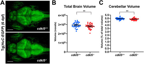
Brain imaging in cdkl5−/− fish. (A) Maximum intensity projections of confocal images from whole brains from Tg(HuC:EGFP); cdkl5+/+ and Tg(HuC:EGFP); cdkl5−/− fish at 6 dpf. Scale bars: 100 µm. (B,C) Quantification of total brain (B) and cerebellar (C) volume from cdkl5+/+ and cdkl5−/− fish. Data are mean±s.e.m. for three independent experiments (n=12, 12 and 11 for cdkl5+/+, and n=12, 12 and 12 for cdkl5−/− fish), **P<0.01 (two-tailed t-test). 1 voxel=8 µm3.

EXPRESSION / LABELING:
Gene:
Fish:
Anatomical Term:
Stage: Day 6
PHENOTYPE:
Fish:
Observed In:
Stage: Day 6

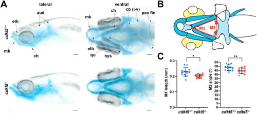
Alcian Blue staining of craniofacial cartilage structures in cdkl5−/− fish. (A) Dorsal and ventral images of Alcian Blue staining in cdkl5+/+ and cdkl5−/− fish at 7 dpf. aud, auditory capsule; cb, ceratobranchials or gill arch derivatives; ch, ceratohyal; eth, ethmoid plate; hys, hyosymplectic; mk, Meckel's cartilage; pec fin, pectoral fins; qu, quadrate. Scale bars: 100 µm. (B) Schematic of Alcian Blue staining in zebrafish heads at 7 dpf, showing locations of head measurements. (C) Quantification of the length (M1) and intersecting angle (M2) of the ceratohyal cartilage structures in cdkl5+/+ and cdkl5−/− fish at 7 dpf. Data are mean±s.d. (n=13 fish per genotype), *P<0.05, **P<0.01 (two-tailed t-test).

ZFIN is incorporating published figure images and captions as part of an ongoing project. Figures from some publications have not yet been curated, or are not available for display because of copyright restrictions.

ZFIN is incorporating published figure images and captions as part of an ongoing project. Figures from some publications have not yet been curated, or are not available for display because of copyright restrictions.

Acknowledgments
This image is the copyrighted work of the attributed author or publisher, and ZFIN has permission only to display this image to its users. Additional permissions should be obtained from the applicable author or publisher of the image. Full text @ Dis. Model. Mech.