
Figure 3
- ID
- ZDB-FIG-210825-41
- Publication
- Dray et al., 2021 - Dynamic spatiotemporal coordination of neural stem cell fate decisions occurs through local feedback in the adult vertebrate brain
- Other Figures
- All Figure Page
- Back to All Figure Page
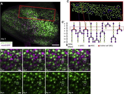
Intravital imaging resolves adult NSC lineage trees in time and space (A) Whole pallial hemisphere imaged intravitally in a 3mpf (B) Close-ups from the same video showing an asymmetric NSC division (dotted circles) between days 3 and 9: 1 daughter differentiates over the next 7 days (bottom dotted circle, loss of the (C and C’) Segmentation and NSC tracking over 23 days in Dm in Mimi. (C) Segmentation of ~390 cells per time point (area boxed in A, at day 6). (C’) Example of dividing tracks, with cell states (color-coded) and the spatial position of each tree (see also |