|
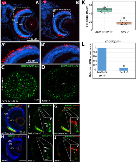
Her9 mutants have fewer rods with abnormal outer segments. Immunohistochemistry with a rod antibody (4C12) in her9+/+ or +/− (A,A′) and her9−/− (B,B′) retinal sections. (C–D) Confocal images of whole eyes from her9 heterozygous incross progeny on the XOPS:GFP background. (E–G) Immunohistochemistry with an antibody that labels rhodopsin (1D1) on retinal cryosections of XOPS:GFP WT and heterozygous (E–G) or her9 mutant (H–J) larvae. (K) Rod cell counts in her9−/− larvae and their WT and heterozygous siblings (# of rods/100 µm). WT/Het = 10 embryos; Mut = 10 embryos; t-test (p < .0001). (L) qPCR analysis of rhodopsin expression at 8 dpf (fold change relative to Ef1α). ONL, outer nuclear layer; OPL, outer plexiform layer; INL, inner nuclear layer; GCL, ganglion cell layer; L, lens; ON, optic nerve; P, pineal gland. Scale bar = 50 µm and 100 µm.
|