
Fig. S3
- ID
- ZDB-FIG-160831-15
- Publication
- Xu et al., 2016 - Microglia Colonization of Developing Zebrafish Midbrain Is Promoted by Apoptotic Neuron and Lysophosphatidylcholine
- Other Figures
- All Figure Page
- Back to All Figure Page
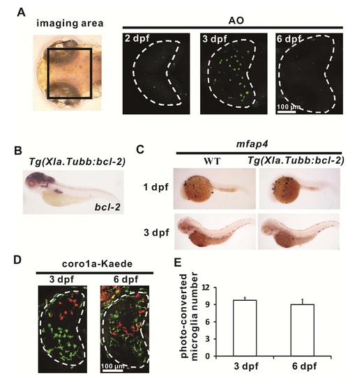
Colonization of the Optic Tectum by Microglial Precursor Depends on Apoptotic Neurons, related to Figure 3 (A) Acriding Orange (AO) staining (green) reveals the accumulation of apoptotic neurons in the optic tectum of the zebrafish brain from 2 dpf to 3 dpf and the subsequent reduction of apoptotic neurons at 6 dpf. The optic tectum is indicated by dashed lines. (B) WISH shows a robust expression of bcl-2 in the brain of the 3 dpf Tg(Xla.Tubb:bcl-2) embryos. (C) mfap4 WISH staining shows normal development of peripheral macrophages in Tg(Xla.Tubb:bcl-2) embryo during early development. (D) Photo-converted coro1a-Kaede+ microglia (red signals) in the optic tectum (white dashed lines) at 3 dpf and 6 dpf . (E) Quantification of Photo-converted coro1a-Kaede+ microglia at 3 dpf and 6 dpf . n=4 for 3 dpf and 6 dpf embryos. Error bars represent mean SEM. |
Reprinted from Developmental Cell, 38(2), Xu, J., Wang, T., Wu, Y., Jin, W., Wen, Z., Microglia Colonization of Developing Zebrafish Midbrain Is Promoted by Apoptotic Neuron and Lysophosphatidylcholine, 214-22, Copyright (2016) with permission from Elsevier. Full text @ Dev. Cell