
|
Fig. S3
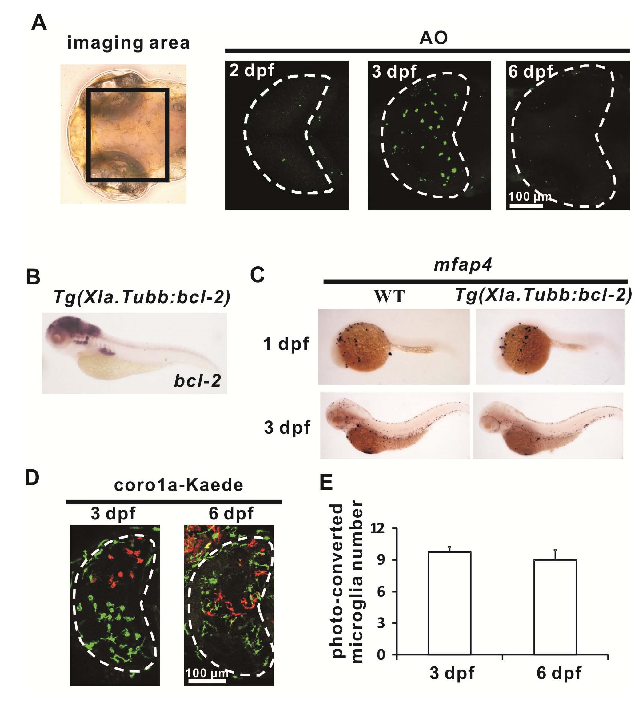
Colonization of the Optic Tectum by Microglial Precursor Depends on Apoptotic Neurons, related to Figure 3
(A) Acriding Orange (AO) staining (green) reveals the accumulation of apoptotic neurons in the optic tectum of the zebrafish brain from 2 dpf to 3 dpf and the subsequent reduction of apoptotic neurons at 6 dpf. The optic tectum is indicated by dashed lines.
(B) WISH shows a robust expression of bcl-2 in the brain of the 3 dpf Tg(Xla.Tubb:bcl-2) embryos.
(C) mfap4 WISH staining shows normal development of peripheral macrophages in Tg(Xla.Tubb:bcl-2) embryo during early development.
(D) Photo-converted coro1a-Kaede+ microglia (red signals) in the optic tectum (white dashed lines) at 3 dpf and 6 dpf .
(E) Quantification of Photo-converted coro1a-Kaede+ microglia at 3 dpf and 6 dpf . n=4 for 3 dpf and 6 dpf embryos. Error bars represent mean SEM.
Reprinted from Developmental Cell, 38(2), Xu, J., Wang, T., Wu, Y., Jin, W., Wen, Z., Microglia Colonization of Developing Zebrafish Midbrain Is Promoted by Apoptotic Neuron and Lysophosphatidylcholine, 214-22, Copyright (2016) with permission from Elsevier. Full text @ Dev. Cell