
Fig. S2
- ID
- ZDB-FIG-160831-14
- Publication
- Xu et al., 2016 - Microglia Colonization of Developing Zebrafish Midbrain Is Promoted by Apoptotic Neuron and Lysophosphatidylcholine
- Other Figures
- All Figure Page
- Back to All Figure Page
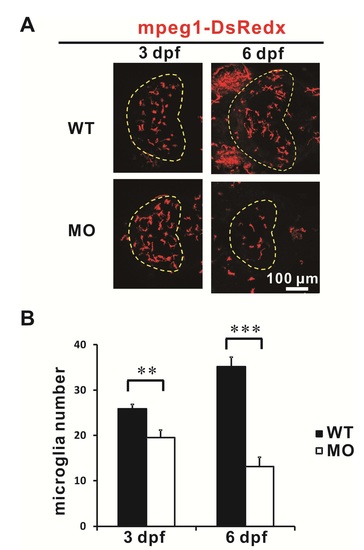
Microglial Precursors Enter the Optic Tectum via Non-circulation route, related to Figure 2 (A) Dorsal view of the optic tectum of Tg(mpeg1:loxP-DsRedx-loxP-GFP) zebrafish embryos injected with or without the tnnt2a morpholno. The number of the optic tectum-resident microglia only slightly decreased in the 3 dpf tnnt2a morphants (MO) but is drastically reduced in the 6 dpf tnnt2a morphants. The optic tectum is indicated by dashed lines. (B) Quantification of the optic tectum-resident microglia number in WT embryos and tnnt2a morphants. Error bars represent mean SEM. **: p<0.01. ***: p<0.001. (n=8 for both WT and morphants at 3 dpf and 6 dpf) |
Reprinted from Developmental Cell, 38(2), Xu, J., Wang, T., Wu, Y., Jin, W., Wen, Z., Microglia Colonization of Developing Zebrafish Midbrain Is Promoted by Apoptotic Neuron and Lysophosphatidylcholine, 214-22, Copyright (2016) with permission from Elsevier. Full text @ Dev. Cell