
Fig. 5
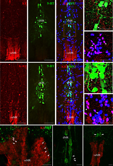
Neural association of kisspeptin 1 (Kiss1) and Kiss-R1 fibers (red) with serotonergic (green) and non-serotonergic (blue) raphe neurons in pet1 Tg zebrafish. (a–c) and F-H: Kiss1 (a–c) and Kiss-R1 (f–h) fiber terminal was seen in the vaMR, while pet1-green fluorescent protein (GFP) labeled 5-HT neurons were noted in the MR. (d, e, i and j) Confocal image of double-labeling showed close association of Kiss1 (d and e) and Kiss-R1 (i and j) fibers with 5-HT neurons in the dMR (d and i) and non-5-HT cells (DAPI) in the vaMR (e and j). There was no co-expression of Kiss-R1 in 5-HT cells. (60× plus 1.5× optical zoom; N.A. = 1.4; z-step = 0.15 µm). (k–m) 5-HT fibers were seen in close association with Kiss1 fibers in the vaMR (k) and in the dMR (l). There were few 5-HT cells seen near the vaMR region, but not inside the vMR (m). Scale bars, (a–c) and (f–g) 100 µm; (d, e, i and j) 20 µm; (k–m) 10 µm. |