
FIGURE
Fig. 2
Fig. 2
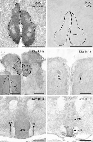
Expression of kissr1 mRNA and Kiss-R1 projection in the zebrafish brain. Coronal sections where kissr1 mRNA is noted in the ventral subnuclei of the habenula (a) and no cells were observed in the sense strand (b). Kiss-R1-immunoreactive (-ir) cells observed in the vHb (c) send projections through the fasciculus retroflexus (FR) (d and e) down to the vaMR (f). Preabsorption with antigen showed no Kiss-R1–ir fibers or cells (C inset). Scale bars, 100 µm. |
Expression Data
Expression Detail
Antibody Labeling
Phenotype Data
Phenotype Detail
Acknowledgments
This image is the copyrighted work of the attributed author or publisher, and
ZFIN has permission only to display this image to its users.
Additional permissions should be obtained from the applicable author or publisher of the image.
Full text @ J. Neurochem.