|
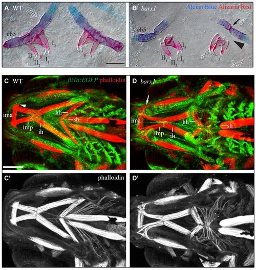
barx1 mutant zebrafish display tooth and muscle phenotypes. (A,B) 5 dpf Alcian Blue- and Alizarin Red-stained ceratobranchial 5 (cb5) and pharyngeal teeth (annotated I1-II2) were dissected and imaged with transmitted light. Arrow indicates ectopic joint in cb5. Arrowhead illustrates missing bone of attachment. Ventral view, anterior is upwards, right is towards the left. Scale bar: 50 μm. (C,D) Confocal projections of 5 dpf larvae with pharyngeal arch derivatives transgenically labeled with EGFP (fli1a:EGFP, green) and muscles stained with phalloidin (red). Ventral view, anterior is towards the left, left is upwards. Labeled muscles include the intermandibularis posterior (imp), intermandibularis anterior (ima), interhyoideus (ih), hyohyoideus (hh) and sternohyoideus (sh). Arrowhead indicates imp attachment site in wild types, arrow indicates disorganized imp invading the ectopic gap in Meckel’s cartilage in mutants. (C′,D′) Single channel phalloidin fluorescence. Scale bar: 100 μm.
|