
FIGURE
Fig. S3
- ID
- ZDB-FIG-121205-3
- Publication
- Blaker-Lee et al., 2012 - Zebrafish homologs of 16p11.2, a genomic region associated with brain disorders, are active during brain development, and include two deletion dosage sensor genes
- Other Figures
- All Figure Page
- Back to All Figure Page
Fig. S3
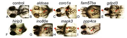
Pigmentation is abnormal in LOF embryos. Pigment cells were observed after LOF, at 48 hpf, by brightfield imaging. Rostral and midline cells were specifically scored as a measure of normal melanocyte migration. (A-I) dorsal views. Genes targeted for LOF by MO injection are indicated above each set of panels. Red arrowhead, no rostral pigment cells; red arrow, no midline pigment cells; scale bar, 150 μM. Average of 35 embryos assayed per gene, over 2-4 independent experiments, 70-100% affected compared to control MO-injected embryos. |
Expression Data
Expression Detail
Antibody Labeling
Phenotype Data
| Fish: | |
|---|---|
| Knockdown Reagents: | |
| Observed In: | |
| Stage: | Long-pec |
Phenotype Detail
Acknowledgments
This image is the copyrighted work of the attributed author or publisher, and
ZFIN has permission only to display this image to its users.
Additional permissions should be obtained from the applicable author or publisher of the image.
Full text @ Dis. Model. Mech.