Fig. 6
- ID
- ZDB-FIG-101021-35
- Publication
- Lyons et al., 2010 - Carboxypeptidase A6 in zebrafish development and implications for VIth cranial nerve pathfinding
- Other Figures
- All Figure Page
- Back to All Figure Page
|
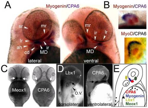
Distribution of CPA6 mRNA compared with tissue-specific markers at 2 dpf. (A) A general marker of muscle precursors, myogenin [16], labels most extraocular muscles (orange) by in situ hybridization, but does not co-localize with CPA6 mRNA (purple). (B) Myogenin mRNA, as well as MyoD mRNA, is also found in the pectoral musculature, unlike CPA6 mRNA which is ectodermal. CPA6 mRNA does not co-localize with Meox1 (C), or Lbx1 (D), both putative markers of the lateral rectus muscle in the chick, but likely not in the zebrafish. (E) A schematic of a 2 dpf zebrafish indicates the relative spatial expression of CPA6, myogenin (in lateral rectus), Lbx1 and Meox1. lr, lateral rectus; mr, medial rectus; sr, superior rectus; ah, adductor hyoideus; cd, constrictor dorsalis; MD, myodome precursors; O.V., otic vesicle. |
| Genes: | |
|---|---|
| Fish: | |
| Anatomical Terms: | |
| Stage: | Long-pec |

