
Fig. S2
- ID
- ZDB-FIG-101011-25
- Publication
- Erickson et al., 2010 - Meis1 specifies positional information in the retina and tectum to organize the zebrafish visual system
- Other Figures
- All Figure Page
- Back to All Figure Page
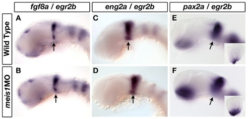
Meis1-knockdown does not affect patterning of the midbrain-hindbrain boundary. (A-F) mRNA in situ hybridization for midbrain-hindbrain boundary (MHB) markers fgf8a (A, B), eng2a (C, D) and pax2a (E, F) in 32-hpf wild-type and meis1 morphant embryos. Arrows indicate the relevant gene expression domain at the MHB. The insets in (E, F) are representative dissected eyes showing an upregulation of pax2a staining in the optic stalk of meis1 morphants (n = 18/18). Embryos are co-stained with the hindbrain r3 and r5 marker egr2b. Embryos are shown in lateral view with dorsal up and anterior to the left, and the dissected retinas are oriented with dorsal up and nasal to the left. |