|
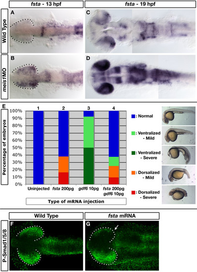
follistatin a is ectopically expressed in Meis1-depleted embryos and can inhibit Gdf6-mediated Bmp signalling. (A-D) mRNA in situ hybridizations for fsta on 13-hpf (A, B) and 19-hpf (C, D) wild-type and meis1 morphant (meis1MO) embryos. Dotted lines outline the optic vesicle. All views are dorsal with anterior to the left. (E) Results of the GDF6-Fsta interaction experiments. One-cell embryos were injected with either 200 pg of zebrafish fsta mRNA (bar 2), human GDF6 mRNA (bar 3), or both mRNAs (bar 4), raised until 28 hpf, and scored for dorsalized and ventralized phenotypes (see legend on the right for classification). (F, G) Confocal images of whole mount immunostains for phospho-Smad1/5/8 in wild-type and fsta mRNA-injected embryos at 14 hpf. Injection of fsta mRNA into one cell of a two-cell embryo causes a unilateral reduction in phospho-Smad1/5/8 staining (arrow in G). Dotted lines outline the optic vesicle. Views are dorsal with anterior to the left.
|

