Fig. 1
- ID
- ZDB-FIG-080424-16
- Publication
- Whitlock et al., 2003 - Gonadotropin-releasing hormone (gnrh) cells arise from cranial neural crest and adenohypophyseal regions of the neural plate in the zebrafish, Danio rerio
- Other Figures
- All Figure Page
- Back to All Figure Page
|
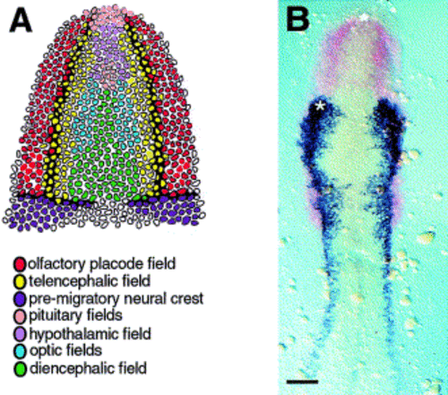
The olfactory placode field abuts the CNC and anterior pituitary fields. (A) Summary fate map incorporating olfactory placode, telencephalic field and edge of CNC (Whitlock and Westerfield, 2000), and eye/diencephalic field (Varga et al., 1999) from zebrafish. Pituitary and hypothalamic domains are based on chick fate map (Couly and Le Douarin 1987 and Le Douarin and Kalcheim 1999). (B) Double in situ hybridization for dlx3 (Akimenko et al., 1994) (red) in olfactory field and fkh6 (Odenthal and Nusslein-Volhard, 1998) (blue) in premigratory CNC. The expression of dlx3 marks the developing olfactory field and otic placode field. There is a row of cells expressing dlx3 that runs along the fkh6 domain. Asterisk marks area where Dil was applied in the living embryo; the star marks the adenohypophyseal region. Scale bar in (B), 100 μm |
Reprinted from Developmental Biology, 257(1), Whitlock, K.E., Wolf, C.D., and Boyce, M.L., Gonadotropin-releasing hormone (gnrh) cells arise from cranial neural crest and adenohypophyseal regions of the neural plate in the zebrafish, Danio rerio, 140-152, Copyright (2003) with permission from Elsevier. Full text @ Dev. Biol.

