
Fig. 7
- ID
- ZDB-FIG-051220-29
- Publication
- Chen et al., 2005 - Loss of function of def selectively up-regulates {Delta}113p53 expression to arrest expansion growth of digestive organs in zebrafish
- Other Figures
- All Figure Page
- Back to All Figure Page
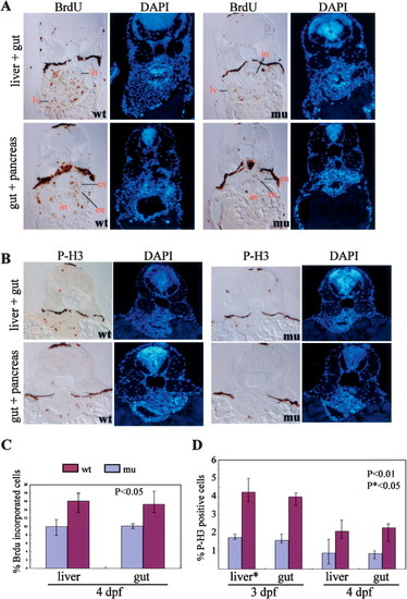
The defhi429 mutant phenotype is caused by compromised cell proliferation. (A,C) BrdU labeling revealed that, at 4 dpf, the number and ratio of cells entering the G1-to-S-phase transition was significantly reduced in the digestive organs of the defhi429 mutant (mu) when compared with the wild type (wt). (Top panel) Cross-sectioning through liver (lv) and intestine (in). (Bottom panel) Cross-sectioning through pancreas and intestine (in). The BrdU incorporation ratios shown in C were obtained by counting BrdU-labeled cells versus total cells in a specific organ (e.g., liver) in sections from seven wild-type and seven mutant embryos, respectively. (en) Endocrine pancreas; (ex) exocrine pancreas. (B,D) At 3 and 4 dpf, histochemical staining using anti-phosphorylated histone 3 (anti-P-H3) revealed that the number and ratio of cells entering G2 to M phase was drastically reduced in the digestive organs of the defhi429 mutant (mu) when compared with the wild type (wt). The ratios of P-H3-positive cells shown in D were obtained by counting P-H3-positive cells versus total cells in a specific organ (e.g., liver) in sections from three wild-type and three mutant embryos, respectively. |