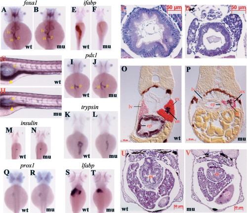
def is required for the expansion growth of intestine, liver, and exocrine pancreas but not endocrine pancreas. (A,B)At2 dpf, whole-mount in situ hybridization using the pan-endoderm marker foxa1 showed that there were no discernible phenotypic differences between wild type (wt) and defhi429 mutant (mu). (in) Intestine; (lv) liver; (p) pancreas. (C,D) At 5 dpf, cross-sectioning showed that the columnar epithelium did form in the mutant gut (mu) but did not fold properly obviously due to greatly reduced cell number when compared with the wild type (wt). (E-H) The expression of gut-specific markers ifabp at 4 dpf (E,F) and alkaline phosphatase at 5 dpf (G,H, yellow arrows) was greatly reduced in the defhi429 mutant. (mu) Mutant; (sw) swimbladder; (wt) wild type. (I,J) At 2 dpf, whole-mount in situ hybridization using a pdx1 probe showed that the initiation and formation of pancreatic bud (p) was not affected in the defhi429 mutant (mu). (in) Intestine. (K,L) At 3 dpf, whole-mount in situ hybridization using a trypsin probe showed that the defhi429 mutant exhibited a much smaller exocrine pancreas. (M,N) In contrast, at 4 dpf, whole-mount in situ hybridization using an insulin probe showed that the endocrine pancreas had no discernible difference between the defhi429 mutant (mu) and the wild type (wt). (O,P) At 4.5 dpf, sectioning in situ hybridization using Fast Red (Roche)-labeled trypsin (for exocrine pancreas, in red) and DIG-labeled insulin (for endocrine pancreas, in purple) probes further confirmed that, when compared with the wild type (wt), the mass of the exocrine pancreas (ex) was greatly reduced while the endocrine pancreas (en) appeared normal in the defhi429 mutant (mu). The mutant epithelium (ep) was narrower and only weakly expressed ifabp (in purple). The liver (lv) in the wild type was labeled using a transferrin probe (for liver, red). (Q,R) At 2 dpf, whole-mount in situ hybridization using prox1 did not reveal a discernible difference between the defhi429 mutant (mu) and the wild type (wt). (S,T) At 4 dpf, the liver bud was obviously smaller and failed to form two lobes in the defhi429 mutant. (U,V) At 5 dpf, cross-sectioning through the esophagus (es) revealed that bile ducts and blood vessels were properly formed in the defhi429 mutant liver (mu) as in the wild-type liver (wt).
|

