
- Title
-
SRPK3 Is Essential for Cognitive and Ocular Development in Humans and Zebrafish, Explaining X-Linked Intellectual Disability
- Authors
- Roychaudhury, A., Lee, Y.R., Choi, T.I., Thomas, M.G., Kahn, T.N., Yousaf, H., Skinner, C., Maconachie, G., Crosier, M., Horak, H., Constantinescu, C.S., Kim, T.Y., Lee, K.H., Kyung, J.J., Wang, T., Ku, B., Chodirker, B.N., Hammer, M.F., Gottlob, I., Norton, W.H.J., Gorlei, R., Kim, H.G., Graziano, C., Pippucci, T., Lovino, E., Montanari, F., Severi, G., Toro, C., Boerkoel, C.F., Cha, H.S., Choi, C.Y., Kim, S., Yoon, J.Y., Gilmore, K., Vora, N.L., Davis, E.E., Chudley, A.E., Schwartz, C.E., and Kim, C.H.
- Source
- Full text @ Ann. Neurol.

Pedigree of Family 1 demonstrating X-linked pattern of inheritance. (A) The first variant in the SRPK3 gene (c.475C > G; p.H159D) was identified in 5 affected males (F1:II-1, F1:III-3, F1:III-4, F1:III-5, F1:III-7) from a well-established X-linked intellectual disability (XLID) family. (B) Photographs of 4 (F1:II-1, F1:III-4, F1:III-5, F1:III-7) affected males. (C) Brain imaging were performed on 4 of the 5 affected males. A representative MRI image of the patient F1:III-4, demonstrating a dilatation of the occipital horns. |

CNS expression of SRPK3 in human and zebrafish embryos. (A) At Carnegie stage 15 (CS15), immunohistochemistry results showing widespread staining of SRPK3 within the hindbrain (hb). (B) At CS19 postmitotic expression is noted in the forebrain (fb), cerebellum (cb) and medulla (m). (C) At CS23 expression is noted in cerebellum. (B′, C′) High magnification images showing intense staining of choroid plexus (cp) epithelium and expression within the cerebellum (cb), medulla (m), and eye. (D) Spatiotemporal expression of srpk3 in developing zebrafish embryo showing transcripts of srpk3 being present at 24 hpf in the brain, heart, and muscles, and in the brain and retinal layers at 72 hpf. Scale bars: 1 mm for (A–C), 100 μm for (D). EXPRESSION / LABELING:
|

Molecular modeling of SRPK3 and subcellular localization of SRPK3 variants in HeLa cells. (A) Side chains of 3 amino acids involved in X-linked intellectual disability (XLID) are indicated as spheres on the SRPK3 structure modeled using AlphaFold. Unstructured loop regions of SRKP3 are omitted for clarity, including residues 1–46 and 310–385. (B) Interior misfolding due to H159D and T458N variations. SRPK3 wild-type (WT) left and the H159 and T458N variant form modeled based the AlphaFold structure prediction are shown together. (C) Structural superposition of the AlphaFold-modeled SRPK3 (green) onto the crystal structure of ICP27 (red)-bound SRPK1 (Navy; PDB code 6FAD). Side chain atoms of SRPK3 Glu529 are presented as spheres. (D) Alteration of protein shape and surface charge due to E529K variation. SRPK3 WT (left) and the E529K variant form (right) are shown as sticks and loops (top) or electrostatic surface representation (bottom). (E) Fluorescence cytochemistry images were obtained to compare the localization of SRPK3 WT and variants. Cell nuclei were stained with DAPI, and localization of GFP-SRPK3 was analyzed by observing GFP fluorescence using confocal microscopy. Scale bars; 20 μm. (F) Western blot analysis of GFP-SRPK3 was conducted to confirm the transfection efficiency of WT and variant plasmids. GAPDH served as a loading control. |
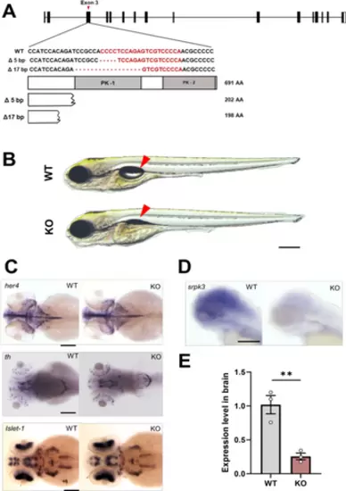
Generation of srpk3 zebrafish knockout (KO) line. (A) Disruption of protein domains in KO zebrafish while wild-type (WT) zebrafish show intact srpk3 protein. Protein kinase domain 1, PK-1; protein kinase domain 2, PK-2. (B) KO zebrafish fail to develop swim bladder inflation at 5 dpf when compared to WT siblings (red arrow). (C) Expression analysis of neuronal markers (her4, th, islet1) and srpk3 at 72hpf. Scale bars; 200 μm. (D, E) Reduced expression level of srpk3 mRNA in KO zebrafish. Three adult brains for each group were used in qRT-PCR experiments (n = 3 for WT and n = 3 for KO). EXPRESSION / LABELING:
PHENOTYPE:
|

Analysis of spontaneous eye movements in knockout (KO) zebrafish. (A) Representative image of spontaneous eye movement in wild-type (WT) and KO zebrafish at 5 dpf. (B) Individual representation of spontaneous eye movement in WT and srpk3 KO siblings. Red lines represent right eye and blue lines represent left eye of 5 dpf zebrafish larvae. (C) Eye movement frequency in the left (L) and right (R) eye of srpk3 KO zebrafish when compared to its WT siblings. (D) Ocular angle significantly decreases in both left and right eye in KO zebrafish, compared to WT siblings. (E) srpk3 KO zebrafish take significant longer time to reset in slow phase when compared to their WT siblings during spontaneous eye movement. Video recording of WT (n = 11) and KO (n = 12) zebrafish for 3 min. All data is represented as mean ± SEM. **p < 0.01, ***p < 0.001 by t test. PHENOTYPE:
|
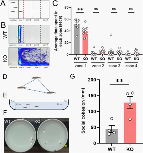
srpk3 knockout (KO) adult zebrafish show impairment in social interaction. (A) Representative image of the experimental setup of social interaction assay in adult zebrafish. (B) Heat map analysis of social interaction assay in adult zebrafish. Three wild-type (WT) fish were used as social cue and 1 as an experimental fish. Black bold line indicates the position of the transparent separator in the water tank. Red and black dashed lines show relative positions of 4 different zones. Video tracking was analyzed at 2 different time bins; early stage (2–7 min) and late stage (10–15 min), respectively. (C) Mean time spent by the experimental fish in each zone at late stage of social interaction assay. (D) Representative image of 3 shoaling fish. (E) Representative image of the parameters of bowl used for shoaling bowl assay. (F) Experimental set up for shoaling bowl assay. Individual adult fish were pointed by arrows. (G) Quantified data for the average of the inter-fish distance of the shoaling assay. The test was performed in triplicates. For each trial, 3 fish of each genotype were used. Number of experimental fish used; n = 10 for WT and n = 10 for KO. All data are represented as mean ± SEM. **p<0.01 by t test. PHENOTYPE:
|
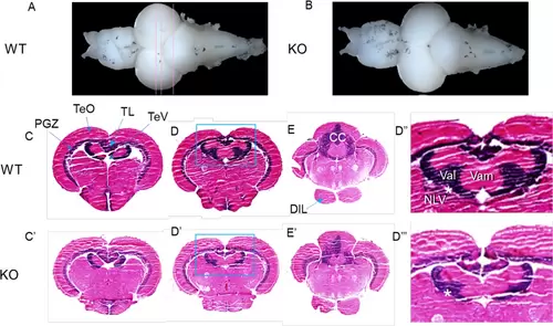
Reduced size of the valvular cerebelli in adult brain of srpk3 KO zebrafish. (A, B) Representative whole brains dissected from a control wild-type (WT) and a knockout (KO) zebrafish. Doral view, anterior is to the left. (C, D‴) Anatomical analysis of brain sections after H&E stain. Frontal sections at 3 different levels indicated in (A). (D″–D‴) Enlargement of valvular cerebelli region in the inlets in (D) and (D′). Asterisk indicates a structural connection between the nucleus lateralis valvulae (NLV) and the valvular cerebelli. Number of adult zebrafish brain used for analysis; n = 3 for WT and n = 3 for KO. CC = corpus cerebelli; DIL = diffuse nucleus of the inferior lobe; NLV = nucleus lateralis valvulae; PGZ = periventricular gray zone of optic tectum; TeO = tectum opticum; TeV = tectal ventricle; TL = torus longitudinalis; Val = lateral division of valvular cerebelli; Vam = medial division of valvular cerebelli. PHENOTYPE:
|