- Title
-
C24:0 avoids cold exposure-induced oxidative stress and fatty acid β-oxidation damage
- Authors
- Sun, S., Cao, X., Gao, J.
- Source
- Full text @ iScience
|
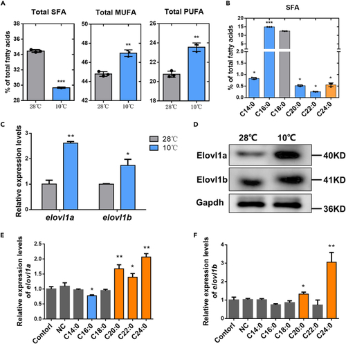
Fatty acid compositions and elovl1/Elovl1 expression levels of zebrafish liver (ZFL) cells (A) The fatty acid compositions of ZFL cells at 28°C and 10°C. (B) The SFA species contents of ZFL cells at 10°C. (C) The mRNA expression levels of elovl1a and elovl1b in ZFL cells at 28°C and 10°C. (D) The protein expression levels of Elovl1a and Elovl1b in ZFL cells at 28°C and 10°C. (E and F) The expression levels of elovl1a (E) and elovl1b (F) in control and SFA-treatment ZFL cells at 28°C. The blue and orange colors in (B), (E), and (F), respectively, meant significant decrease and increase in parameters of treated groups, compared with the control (the control in (B) was the SFA species content of ZFL cells at 28°C). Data were given as means ± SD of three biological replicates. The statistical analyses were conducted by t test. The asterisks labeled above the error bars indicated significant differences (∗p < 0.05, ∗∗p < 0.01, ∗∗∗p < 0.001). SFA, saturated fatty acid; MUFA, monounsaturated fatty acid; PUFA, polyunsaturated fatty acid; NC, negative control; elovl1, fatty acyl elongase 1; Gapdh, glyceraldehyde-3-phosphate dehydrogenase. |
|
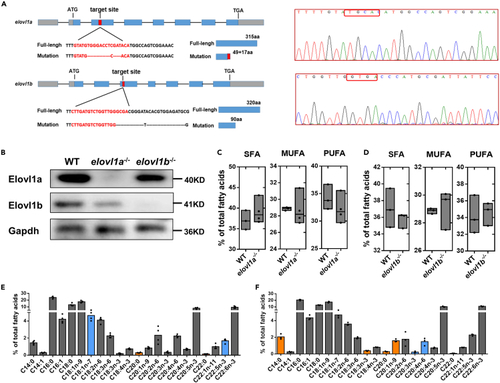
Deletion of elovl1a and elovl1b genes in zebrafish and their effects on fatty acid compositions (A) The targeting site for elovl1a and elovl1b gene knockout. The gray boxes were the 5′-untranslated and 3′-untranslated regions, and blue boxes the exons. The red boxes indicated the targeting sequences. (B) Mutation confirmation, as shown by the results of the protein expression of Elovl1a and Elovl1b. (C and D) The total SFA, MUFA, and PUFA contents in livers of 2-month-old WT, elovl1a–/– (C) and elovl1b–/– (D) zebrafish. (E and F) Fatty acid compositions in livers of 2-month-old elovl1a–/– (E) and elovl1b–/– (F) zebrafish. The blue and orange colors in (E) and (F), respectively, meant significant decrease and increase in parameters of elovl1a–/–/elovl1b–/– zebrafish, compared with WT zebrafish (p < 0.05). Data were given as means ± SD of three or four biological replicates. The statistical analyses were conducted by t test. WT, wild-type zebrafish; elovl1, fatty acyl elongase 1; elovl1a–/–, elovl1a knockout zebrafish; elovl1b–/–, elovl1b knockout zebrafish; SFA, saturated fatty acid; MUFA, monounsaturated fatty acid; PUFA, polyunsaturated fatty acid; Gapdh, glyceraldehyde-3-phosphate dehydrogenase. |
|
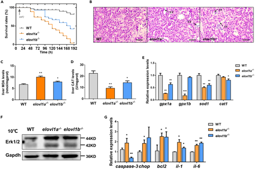
Effects of elovl1a and elovl1b deletion on cold tolerance of zebrafish (A) The survival rates of fish during cold stress. (B) Histological structures of livers from WT, elovl1a–/–, and elovl1b–/– zebrafish under cold stress. Scale bar, 20 μm. (C and D) The levels of MDA (C) and CAT (D) in livers of WT, elovl1a–/–, and elovl1b–/– zebrafish under cold stress (n = 5). (E) Hepatic expression levels of four oxidative stress-related genes in WT, elovl1a–/–, and elovl1b–/– zebrafish under cold stress. (F) Western blotting of Erk1/2 in livers of WT, elovl1a–/–, and elovl1b–/– zebrafish under cold stress. (G) Hepatic expression levels of apoptosis and inflammation-related genes in WT, elovl1a–/–, and elovl1b–/– zebrafish under cold stress. Data were given as means ± SD of three biological replicates unless otherwise specified. The statistical analyses were conducted by t test. The asterisks labeled above the error bars indicated significant differences (∗p < 0.05, ∗∗p < 0.01, ∗∗∗p < 0.001). WT, wild-type zebrafish; elovl1, fatty acyl elongase 1; elovl1a–/–, elovl1a knockout zebrafish; elovl1b–/–, elovl1b knockout zebrafish; LY, lymphocyte; MDA, malondialdehyde; CAT, catalase; gpx-1a, glutathione peroxidase 1a; sod1, superoxide dismutase 1, soluble; cat1, catalase 1; Erk1/2, extracellular signal-regulated kinases 1/2; Gapdh, glyceraldehyde-3-phosphate dehydrogenase; chop, DNA damage inducible transcript 3; bcl2, BCL2 apoptosis regulator a; il, interleukin. |
|
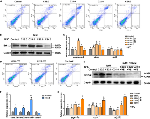
Effects of different saturated fatty acids (SFA, C18–C24) on cold tolerance of zebrafish liver (ZFL) cells (A) Apoptotic levels of ZFL cells under cold stress, after incubation with different SFAs. (B) Western blotting of Erk1/2 in ZFL cells under cold stress, after incubation with different SFAs. (C) The expression levels of apoptosis-related genes in ZFL cells under cold stress, after incubation with different SFAs. (D) Apoptotic levels of ZFL cells under cold stress, after incubation with different SFAs + VE. (E) Western blotting of Erk1/2 in ZFL cells under cold stress, after incubation with different SFAs + VE. (F) The expression levels of four ceramide synthase genes in ZFL cells under cold stress, after incubation with C24:0. (G) The expression levels of β-oxidation-related genes in ZFL cells under cold stress, after incubation with different SFAs. Data were given as means ± SD of three biological replicates. The statistical analyses were conducted by t test or one-way ANOVA with Tukey's post hoc test. The different letters above the bars indicated significant differences (p < 0.05). The asterisks labeled above the error bars indicated significant differences (∗p < 0.05, ∗∗p < 0.01, ∗∗∗p < 0.001). Erk1/2, extracellular signal-regulated kinases 1/2; Gapdh, glyceraldehyde-3-phosphate dehydrogenase; chop, DNA damage inducible transcript 3; bcl2, BCL2 apoptosis regulator a; VE, vitamin E; cers, ceramide synthase; pgc-1a, peroxisome proliferator-activated receptor-γ coactivator-1a; cpt-1a, carnitine palmitoyl transferase 1a; atp5b, ATP synthase F1 subunit beta. |
|
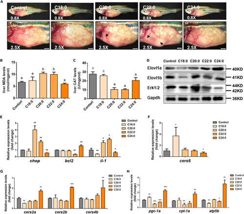
Effects of different dietary saturated fatty acids (SFA) on cold tolerance of zebrafish (A) The liver lesions of zebrafish under cold stress, after being fed with different SFA diets for 2 weeks. Scale bar, 1 mm. (B and C) The levels of hepatic MDA (B) and CAT (C) in zebrafish under cold stress, after being fed with different SFA diets for 2 weeks. (D) Western blotting of Elovl1a, Elovl1b, and Erk1/2 in livers of zebrafish under cold stress, after being fed with different SFA diets for 2 weeks. (E) Hepatic expression levels of apoptosis and inflammation-related genes in zebrafish under cold stress, after being fed with different SFA diets for 2 weeks. (F and G) The expression levels of four ceramide synthase genes in livers of zebrafish under cold stress, after being fed with different SFA diets for 2 weeks. (H) The expression levels of β-oxidation-related genes in livers of zebrafish under cold stress, after being fed with different SFA diets for 2 weeks. Data were given as means ± SD of three biological replicates. The statistical analyses were conducted by t test or one-way ANOVA with Tukey's post hoc test. The different letters above the bars indicated significant differences (p < 0.05). The asterisks labeled above the error bars indicated significant differences (∗p < 0.05, ∗∗p < 0.01, ∗∗∗p < 0.001). MDA, malondialdehyde; CAT, catalase; Elovl1, fatty acyl elongase 1; Erk1/2, extracellular signal-regulated kinases 1/2; Gapdh, glyceraldehyde-3-phosphate dehydrogenase; chop, DNA damage inducible transcript 3; bcl2, BCL2 apoptosis regulator a; il, interleukin; cers, ceramide synthase; pgc-1a, peroxisome proliferator-activated receptor-γ coactivator-1a; cpt-1a, carnitine palmitoyl transferase 1a; atp5b, ATP synthase F1 subunit beta. |

