FIGURE SUMMARY
Title

Developmental Angiogenesis Requires the Mitochondrial Phenylalanyl-tRNA Synthetase

Authors
Li, B., Chen, K., Liu, F., Zhang, J., Chen, X., Chen, T., Chen, Q., Yao, Y., Hu, W., Wang, L., Wu, Y.
Source
Full text @ Front Cardiovasc Med

The expression of fars2 is essential in the early stage of zebrafish embryo development. (A) The expression patterns of fars2 during zebrafish embryonic development. The qRT-PCR analyses were performed at eight embryo development stages (6, 24, 30, 48, 72, 96, 120, and 144 hpf). (B) The percentages of embryos with developmental defects in zebrafish injected with a non-specific control or fars2-specific morpholinos (MOs). (C) Representative images of zebrafish at 50 hpf, following an injection with a non-specific control or fars2-specific MOs. ***P < 0.001.

EXPRESSION / LABELING:
Gene:
Fish:
Anatomical Term:
Stage Range: Shield to Day 6

The morpholino-mediated knock-down of fars2 delays vascular formation in zebrafish. (A) Representative images of the trunk regions of Tg (fli1: EGFP)y1 embryos taken at 50 hpf. The intersegmental vessel (white arrow), dorsal longitudinal anastomotic vessel (blue arrow), parachordal vessel (red arrow), and ectopic sprouts (asterisks) are indicated. (B,C) The number of complete intersegmental vessels (ISVs) and the mean lengths of the ISVs in control and fars2 morphants. The horizontal bars show the mean ± SEM (n = 10 per group). ***P < 0.001 via ANOVA. (D) Representative images of the caudal artery, caudal vein, and caudal vein plexus (CVP; arrows) in Tg (fli1: EGFP)y1 embryos taken at 50 hpf. In the control embryo, the CVP formed a typical honeycomb structure in the tail (white arrows). The knock-down of fars2 resulted in specific defects in CVP formation (yellow arrows). (E) Quantification of the loop number at the CVP. The horizontal bars show the mean ± SEM (n = 10 per group). ***P < 0.001 via ANOVA.

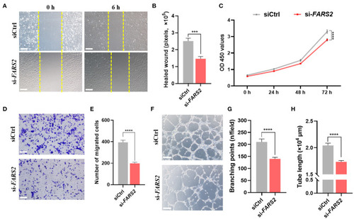
The deficiency of FARS2 impairs cell motility, proliferation, invasion, and tube formation in human umbilical vein endothelial cells (HUVECs). (A) Representative images of scratch-wound assays of HUVECs 0 and 6 h at 48 h after transfection with a control (siCtrl) or FARS2-specific (si-FARS2) siRNA. Scale bar = 200 μm. (B) Quantification of the healed wound area from (A). Data are prepresented as the mean and SEM (n = 10). ***P < 0.001 via ANOVA. (C) A CCK8-based cell proliferation assay of HUVECs at the indicated time-points after transfection with siCtrl or si-FARS2. The measurements were made in triplicate (mean and SEM), and the results are indicative of three independent experiments. ****P < 0.0001. (D) Representative images of transwell-based migration assays of HUVECs 48 h after transfection with siCtrl or si-FARS2. Scale bar = 200 μm. (E) Quantification of the number of migrated cells from (D). ***P < 0.001. (F) Representative images of tube network assays of HUVECs 48 h after transfection with siCtrl or si-FARS2. Scale bar = 200 μm. (G,H) Quantification of the branching points (G) and tube lengths (H) from (F). The measurements were made in triplicate (mean and SEM), and the results are indicative of three independent experiments. ****P < 0.0001.

FARS2 silencing causes mitochondrial dysfunction in human umbilical vein endothelial cells (HUVECs). (A) The oxygen consumption rate (OCR) in HUVECs transfected with siCtrl or si-FARS2. The HUVECs were seeded 48 h after transfection with siRNAs and 12 h before analysis using a Seahorse XF24 analyzer. The OCR was measured continuously throughout the experimental period, both at baseline and in the presence of the indicated drugs. (B) Non-mitochondrial respiration, basal respiration, maximal respiration, proton leak, ATP production, and spare respiratory capacity in control and FARS2-deficient HUVECs. The measurements were made in triplicate (mean and SEM). **P < 0.01, ***P < 0.001, ****P < 0.0001. (C) The effects of FARS2 knock-down on intracellular reactive oxygen species production by HUVECs. The measurements were made in triplicate (mean and SEM). *P < 0.05, (D) Quantification of total ATP levels in HUVECs 48 h after transfection with the indicated siRNAs. The measurements were made in triplicate (mean and SEM). **P < 0.01.

The deficiency of FARS2 impairs angiogenesis by disrupting the Notch and Wnt signaling pathways. (A) The expression levels of genes involved in the Notch/Wnt pathways in control and fars2 zebrafish morphants, as determined by qRT-PCR analyses (n = 6–10 individual embryos). ***P < 0.001, **P < 0.01, *P < 0.05; ns, not significant. (B) The relative mRNA expression levels of Notch/Wnt pathway-related genes. The human umbilical vein endothelial cells (HUVECs) were transfected with the indicated siRNAs for 48 h and then harvested for qRT-PCR analysis. The measurements were made in triplicate (mean and SEM), and the results are indicative of three independent experiments. *P < 0.05, **P < 0.01, ***P < 0.001, ****P < 0.0001. (C) Western blot analyses of NOTCH1 and β-catenin protein levels. The HUVECs were transfected with the indicated siRNAs for 48 h prior to analysis. (D) Quantification of the western blotting data described in (C). The measurements were made in triplicate (mean and SEM). *P < 0.05, **P < 0.01.

EXPRESSION / LABELING:
Genes:
Fish:
Knockdown Reagent:
Anatomical Term:
Stage: Long-pec
PHENOTYPE:
Fish:
Knockdown Reagent:
Observed In:
Stage: Long-pec

Developmental angiogenesis requires the mitochondrial phenylalanyl-tRNA synthetase. An overview of the mechanisms by which the deficiency of mitochondrial phenylalanyl-tRNA synthetase impairs angiogenesis by disrupting the Notch/Wnt pathways in zebrafish and human umbilical vein endothelial cells.

Acknowledgments
This image is the copyrighted work of the attributed author or publisher, and ZFIN has permission only to display this image to its users. Additional permissions should be obtained from the applicable author or publisher of the image. Full text @ Front Cardiovasc Med