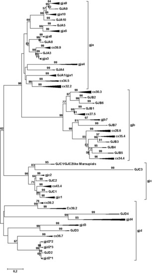
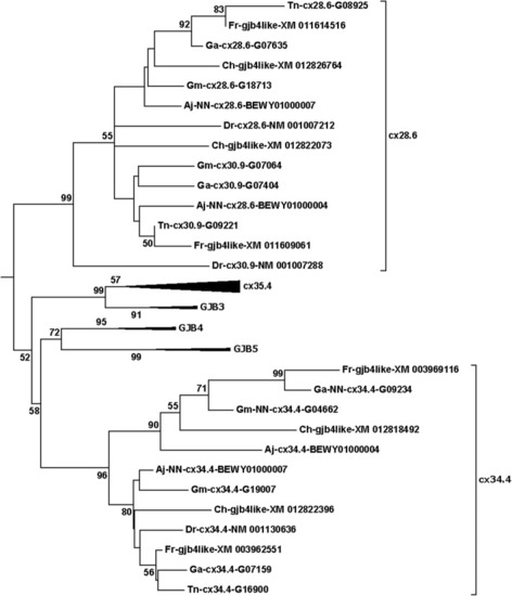
Phylogenetic tree for the gap junction protein (connexin) gene family. The mammalian branches are indicated by upper case letters; teleost branches are indicated by lower case letters. The width of the triangles indicates the number of taxa included in the branch, and the length of the triangles indicates the sequence variation within the branch. The tree was made by the Minimum Evolution method, using amino acids (354 amino acid sequences with 201 positions in the final dataset) and the Dayhoff substitution matrix. The bootstrap values (500 replicates) > 50% are shown next to the branches. To avoid disruptive long-branch attraction, some sequences were excluded (see text). This model gives results that are quite close to the majority of results as summed up in Suppl. Table 1, and thus is close to an average tree from all the phylogenetic analyses. The major difference is that the mammalian GJA10 and teleost gja10 have switched places. In the original three, the root of the GJD family splits up in three very close branches, but using the rooting function in the Mega Tree Explorer, they were collected them into one common basal branch. Note the commonly occurring dichotomy with the mammalian sequences in one of the sub-branches and the teleost sequences in the other sub-branch, although some of the teleost groups do not have a mammalian counterpart (and vice versa). The scale bar (lower left) indicates the number of amino acid substitutions per site

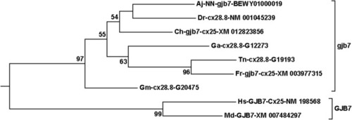
The GJB7/gjb7 branch from the compressed tree shown in Fig. 1. This is an example of a group where all teleost species have only one member, and therefore probably have lost the expected ohnolog partner at a very early stage before the divergence of the different teleosts, similar to most of the other connexins located on the same chromosome (see Table 2). The naming of the sequences is as follows. The two-letter abbreviation indicates the species (Aj, Anguilla japonica = Japanese eel; Dr., Danio rerio = zebrafish; Ch, Clupea harengus = Atlantic herring; Ga, Gasterosteus aculeatus = three-spined stickleback; Tn, Tetraodon nigroviridis = green spotted pufferfish; Fr, Takifugu rubripes = Fugu (Japanese pufferfish); Gm, Gadus morhua = Atlantic cod; Hs, Homo sapiens = human; Md, Monodelphis domestica = opossum) followed by an abbreviation of the name of the sequence in the database (using upper and lower case letters as indicated in the database), and finally, the accession number in the database. NN indicates that there was No Name for the sequence in the database. Further details about the naming are found in the Methods section

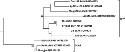
The GJA4/gja4 branch from the compressed tree shown in Fig. 1. This is an example of a group where eel (Aj) has two members, whereas all the other teleosts have one member. The eel pair is found on two different chromosomes (Table 2), suggesting that one member was lost somewhere in-between the divergence of eels and the other teleosts. Moreover, note that the herring (Ch) member is wrongly named gja6like in GenBank; the correct name would be gja4. See legend of Fig. 2 and Methods section for details about naming of the sequences

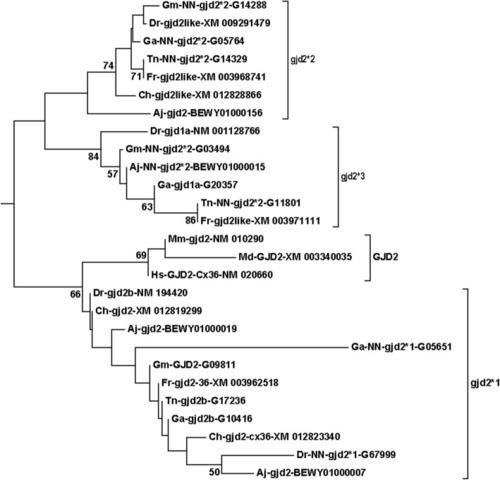
The gjd2 branch from the compressed tree shown in Fig. 1. This is an example of a group where the structure is considerably more complex in teleosts than in mammals. First, there is one teleost group, here called gjd2*1, that in the majority of statistical models locates closest to mammalian GJD2. Gjd2*1 contains two sequences from most fishes, and each members of the pairs are on different chromosomes in all species (Table 2). Secondly, there are two subgroups (here called gjd2*2 and gjd2*3) that are, according to this statistical model, slightly more distantly connected to mammalian GJD2. In this statistical model, the gjd2*2 and gjd2*3 subgroups have a phylogenetic distribution that is “ohnologically perfect” in that it divides into two subgroups containing one sequence from each species. In all species, the pairs of sequences are found on two different chromosomes (Suppl. Table 7). See legend of Fig. 2 and Methods section for details about naming of the sequences

GJB3/GJB4/GJB5 related sequences from the compressed tree shown in Fig. 1. This is an example where teleost sequences with the same names are found in clearly distinct branches of the tree. In this case, four Fugu (Fr) and four herring (Ch) sequences are called gjb4like. Two sequences from each species located into each of the two groups here called cx28.6 and cx34.4. Note also that mammalian GJB4 and GJB5 were always found as a dichotomous pair, and that cx34.4 never mixed into the dichotomous GJB4/GJB5 pair (Suppl. Table 1). Similarly, cx28.6 generally split off at the foot of the collected GJB3/GJB4/GJB5/cx35.5/cx34.4 clade, but in a few cases (with poorer statistics) was positioned closer to GJB3/cx35.4 (Suppl. Table 1). Thus, there is no evidence to support cx28.6 or cx34.4 being more closely related to GJB4 than to GJB5 as the naming (gjb4like) could suggest. See legend of Fig. 2 and Methods section for details about naming of the sequences

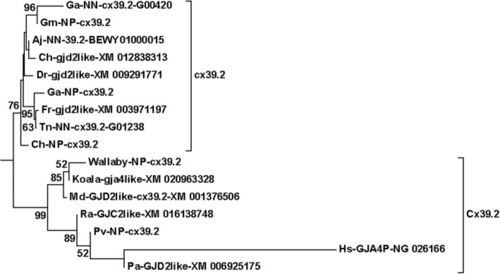
The human pseudogene “GJA4P” (NG_02166) always located together with cx39.2/gjd2like sequences. Note that these “gjd2like” sequences must not be confused with paralogous sequences that have the same name in other groups (cx36.7 and gjd2*2). See legend of Fig. 2 and Methods section for details about naming of the sequences

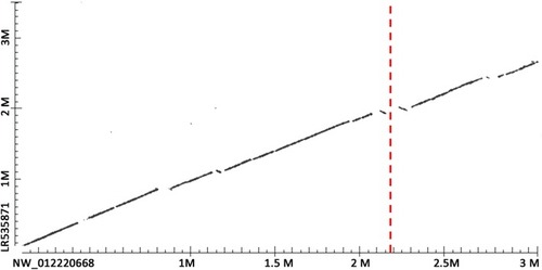
Problem in herring assembly of chromosome 15 at assumed position of gjb7. Scaffold NW_012220668 from the draft herring genome assembly contains gjb7 in position 2,189,757–2,188,978 (i.e., on the reverse strand). This scaffold was aligned with herring chromosome 15 assembly LR535871 position 0 to 3,500,000 using the alignment option in Blast. The position of gjb7 on NW_012220668 is indicated by the red dotted line. There are apparent inversions and breaks in the area where gjb7 was expected in chromosome 15. The word size in the alignment was 256

Acknowledgments
This image is the copyrighted work of the attributed author or publisher, and ZFIN has permission only to display this image to its users. Additional permissions should be obtained from the applicable author or publisher of the image. Full text @ BMC Genomics