FIGURE SUMMARY
Title

A subset of SMN complex members have a specific role in tissue regeneration via ERBB pathway-mediated proliferation

Authors
Pei, W., Xu, L., Chen, Z., Slevin, C.C., Pettie, K.P., Wincovitch, S., NISC Comparative Sequencing Program, Burgess, S.M.
Source
Full text @ NPJ Regen Med

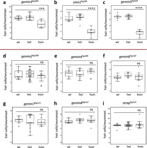
Hair cell regeneration analysis for mutations in each gene of the SMN complex.

Hair cell regeneration is impaired by homozygous mutations of gemin3hg105a, smn1hg104b, and gemin5hg107c, but not in gemin2hg108d, gemin4hg109e, gemin6hg110f, gemin7hg111g, gemin8hg112h, or straphg113i. Red line separates the mutations impacting regeneration from those that have no effect on regeneration. wt wild-type, het heterozygotes, hom homozygotes. Error bars in the graphs represent mean ± s.e.m. The difference between wild-type and homozygote is labeled. ns, P > 0.05; ***P < 0.001; ****P < 0.0001. Approximately 40 embryos were used for each of the regeneration analyses and then genotyped to study the genotype–phenotype correlation.

g<italic>emin5</italic><sup><italic>hg81</italic></sup> mutants possess smaller neuromasts at 2 days post hair cell ablation.

a Live cell imaging of support cells by Tg(tnks1bp1:GFP) and hair cells by Tg(atoh1a:dTomato) in lateral line neuromasts of the gemin5hg81 control and mutant embryos at 2 days post hair cell ablation. Scale bar, 10 µm. b Live cell labeling of neuromast cells by Tg(cldnb:EGFP) in the lateral line of the gemin5hg81 control and mutant embryos at 2 days post hair cell ablation. Dotted white circle demarcates the periphery of the neuromast. Scale bar, 10 µm. c Alkaline phosphatase staining of lateral line neuromasts in the gemin5hg81 control and mutant embryos at 2 days post hair cell ablation. Arrows point to the neuromasts, which are significantly smaller in the mutants when compared to those in the control larvae. Scale bar, 50 µm. d Confocal images of lateral line neuromasts in the gemin5hg81 control and mutant embryos at 2 days post hair cell ablation, stained with anti-hair cell antibodies (green color) and DAPI (blue color). Representative images are shown. Scale bar, 10 µm. e Quantification of neuromast cells. Error bars in the graphs represent mean ± s.e.m. There is a significant reduction in the number of neuromast cells (****P < 0.0001). The numbers are presented as percentage because they were obtained from quantification of still confocal images. Each data point was generated from ~10 embryos.

Decreased neuromast cell proliferation after hair cell ablation in <italic>gemin3</italic>, <italic>gemin5</italic>, and <italic>smn1</italic> mutants.

a Confocal images of lateral line neuromasts in the control and gemin5hg81 mutant at 1-day post hair cell ablation. Neuromasts were stained with DAPI (blue) and proliferating cells were labeled by EdU (pink). The embryos used for the analysis were obtained from a pairwise incross of heterozygotic parents. Hair cells were ablated at 5 dpf. EdU treatment was conducted at 1-day post ablation. Scale bar, 10 µm. b Quantification of EdU-positive cells in the embryos carrying wild-type, heterozygous, or homozygous gemin5hg81 mutations. c Quantification of EdU-positive cells in the embryos carrying wild-type, heterozygous, or homozygous smn1fh229 mutations. d Quantification of EdU-positive cells in the embryos carrying wild type, heterozygous, or homozygous gemin3hg105 mutations. The graphs show mean ± s.e.m. Homozygous mutants for all three regeneration genes have a significantly reduced number of proliferating cells. **P < 0.01; ****P < 0.0001. Data for each mutation were generated using ~40 embryos born from a single pair of heterozygous parents.

<italic>gemin3</italic><sup><italic>hg106</italic></sup> mutations impair regeneration of caudal fins and livers.

a Caudal fin regeneration in the control and gemin3hg106 mutant embryos at 4 days post amputation. Arrows point to the pigment gap which is often missing in the mutants. Scale bar, 100 µm. b Quantification of the area of regenerated caudal fins in the gemin3hg106 mutants. c Liver regeneration in the control and gemin3hg106 mutant embryos at 3 days post ablation. The CFP fluorescence labels the regenerated livers. Scale bar, 100 µm. d Quantification of the area of the regenerated livers. Representative images are shown. Liver tissue is labeled by Tg(fabp10a:CFP-NTR). Graphs show mean ± s.e.m. **P < 0.01; ****P < 0.0001. Data for each analysis were collected from ~40 embryos produced from a pairwise heterozygous incross (for fin regeneration), or a pairwise heterozygous incross in the background of the transgene Tg(fabp10a:CFP-NTR) (for liver regeneration).

Shared genes dysregulated in <italic>gemin3</italic>, <italic>gemin5</italic>, and <italic>smn1</italic> mutants revealed by RNA-Seq.

a Hierarchical clustering of RNA-Seq samples using log2-fold change of normalized read counts. The control and mutant embryos used for the analysis were: smn1fh229 (s1), gemin2hg108 (g2), gemin3hg105 (g3), gemin4hg109 (g4), gemin5hg80 (g5), gemin6hg110 (g6), and gemin7hg111 (g7). The Y-axis shows the linkage value. b and c Log2-fold change of mRNA expression of erbb2b and erbb3bc in different homozygous mutant backgrounds. Error bars indicate standard deviations.

Reduced responsiveness of <italic>gemin3</italic>, <italic>gemin5</italic>, and <italic>smn1</italic> mutants to ErbB pathway inhibitor AG1478.

a Neuromasts in AG1478-treated control and gemin5hg81 mutant embryos at 5 dpf. Neuromasts are shown as white dots. Scale bar, 250 µm. b Quantification of the lateral line neuromasts in mock and AG1478-treated embryos carrying the gemin5hg81 mutation at 5 dpf. c Quantification of the lateral line neuromasts in mock and AG1478-treated embryos carrying the smn1hg104 mutation at 5 dpf. d Quantification of the lateral line neuromasts in mock and AG1478-treated embryos carrying the gemin3hg105 mutation at 5 dpf. e Quantification of the lateral line neuromasts in mock and AG1478-treated embryos carrying the gemin6hg110 mutation at 5 dpf. Error bars in the graphs show the mean ± s.e.m. ns, P > 0.05; *P < 0.05; **P < 0.01; ****P < 0.0001. f Fluorescent images of lateral line neuromasts labeled by transgenes Tg(pou4f3:GAP-GFP) and Tg(SqET20:EGFP) in the control and gemin5hg81 mutant at 5 dpf after AG1478 treatment. Images were taken in the areas surrounding the end of yolk extension. White arrow points to the Tg(SqET20:EGFP) signal in the control embryo, which is dramatically increased in the mutant. Scale bar, 50 µm. The embryos used for the above analyses were generated from a pairwise incross of heterozygotic parents, treated with 2 µM AG1478 from 1 to 5 dpf, and then used Yopro-1 staining or transgenic fluorescence at 5 dpf to analyze neuromast formation. Data for each condition were generated using ~40 embryos born from a single pair of heterozygous parents.

Reduced responsiveness of <italic>gemin5</italic><sup><italic>hg81</italic></sup> mutants to the knockdown of ErbB pathway genes.

a Quantification of lateral line neuromasts in the mock and AG1478-treated erbb3bhg115 mutant embryos at 5 dpf. b Quantification of lateral line neuromasts in the mock and AG1478-treated nrg1hg114 mutant embryos at 5 dpf. Approximately 40 embryos generated from a pair of heterozygous parents carrying either erbb3bhg115 or nrg1hg114 mutation were treated with 0 or 2 µM AG1478 from 1 to 5 dpf and then stained with Yopro-1 to count lateral line neuromasts. c Quantification of lateral line neuromasts in the gemin5hg81/erbb3bhg115 mutant embryos at 5 dpf. The data are generated from analyzing 177 embryos generated from pairwise incrosses of double heterozygous parents and five embryos are double mutants. d Quantification of lateral line neuromasts in the gemin5hg81/nrg1hg114 mutant embryos at 5 dpf. The data are generated from analyzing 156 embryos generated from pairwise incrosses of double heterozygous parents and 13 embryos are double mutants. eerbb2 mutation rate in erbb2 CRISPR guide RNA injected gemin5 mutant embryos at 5 dpf. Mutation rate was measured by CRISPR-STAT fluorescent PCR-based fragment analysis61. f Quantification of lateral line neuromasts in the mock and erbb2 CRISPR guide RNA injected gemin5 mutant embryos at 5 dpf. Error bars in the graphs indicate mean ± s.e.m. ns, P > 0.05; *P < 0.05; **P < 0.01; ****P < 0.0001. The analysis was done by injecting the embryos generated from a pair of gemin5hg81 heterozygous parents with either Cas9 protein alone (mock injection) or Cas9 protein together with erbb2 guide RNAs (erbb2 gRNA injection), followed by analyzing hair cell development in ~40 injected embryos for each condition, and lastly determining gemin5hg81 genotype and erbb2 mutation rate for each of the analyzed embryo. No erbb2 mutation was detected in the mock injection group.

ErbB pathway inhibitor AG1478 partially rescues the hair cell regeneration deficiency in <italic>gemin3</italic>, <italic>gemin5</italic>, and <italic>smn1</italic> mutants.

a Quantification of regenerated hair cells in the AG1478-treated smn1hg104 mutant embryos at 2 days post hair cell ablation. b Quantification of regenerated hair cells in AG1478-treated gemin3hg105 mutant embryos at 2 days post hair cell ablation. c Quantification of regenerated hair cells in lateral line neuromasts in AG1478-treated gemin5hg81 mutant embryos at 2 days post hair cell ablation. The slight reduction in the hair cells of the gemin3hg105 heterozygotes treated with 5 µM of AG1478 could be due to drug toxicity to this genetic background, since it was not observed in the smn1hg104 and gemin5hg81 embryos. Graphs show the mean ± s.e.m. Statistical difference are indicated as: ns, P > 0.05; *P < 0.05; **P < 0.01; ***P < 0.001. Analysis for each condition was done with ~40 embryos generated from a single pair of heterozygous carrier parents, ablated hair cells at 5 dpf, and then treated with 0, 2.5, or 5 µM of AG1478 from 5 to 7 dpf.

ZFIN is incorporating published figure images and captions as part of an ongoing project. Figures from some publications have not yet been curated, or are not available for display because of copyright restrictions.

EXPRESSION / LABELING:
Genes:
Fish:
Anatomical Terms:
Stage Range: Sphere to Days 30-44

ZFIN is incorporating published figure images and captions as part of an ongoing project. Figures from some publications have not yet been curated, or are not available for display because of copyright restrictions.

ZFIN is incorporating published figure images and captions as part of an ongoing project. Figures from some publications have not yet been curated, or are not available for display because of copyright restrictions.

ZFIN is incorporating published figure images and captions as part of an ongoing project. Figures from some publications have not yet been curated, or are not available for display because of copyright restrictions.

Acknowledgments
This image is the copyrighted work of the attributed author or publisher, and ZFIN has permission only to display this image to its users. Additional permissions should be obtained from the applicable author or publisher of the image. Full text @ NPJ Regen Med