FIGURE SUMMARY
Title

Abnormal development of zebrafish after knockout and knockdown of ribosomal protein L10a

Authors
Palasin, K., Uechi, T., Yoshihama, M., Srisowanna, N., Choijookhuu, N., Hishikawa, Y., Kenmochi, N., Chotigeat, W.
Source
Full text @ Sci. Rep.

ZFIN is incorporating published figure images and captions as part of an ongoing project. Figures from some publications have not yet been curated, or are not available for display because of copyright restrictions.

ZFIN is incorporating published figure images and captions as part of an ongoing project. Figures from some publications have not yet been curated, or are not available for display because of copyright restrictions.

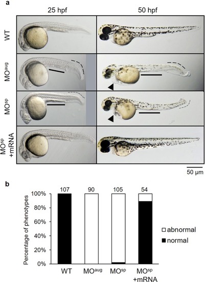
(a) Effect of Rpl10a deficiency on embryonic development. Lateral views of rpl10a MO-injected embryos and rescue phenotype observed at 25 and 50 hpf. At 25 and 50 hpf, smaller eyes, yolk sac extensions, bent tails, and developmental delays were observed in rpl10a knockdown. At 50 hpf, deficient embryos displayed smaller eyes with reduced pigmentation and edema (arrowheads). The MOsp + mRNA injected embryos displayed rescue phenotypes. (b) The phenotype of rpl10a MOaug (n = 90), MOsp (n = 105), and MOsp + mRNA (n = 54) injected embryos was evaluated compared to wild-type (n = 107) at 25 hpf. The morphologic abnormalities include a thinner yolk sac extension, a shorter body length, and a bent tail, whereas the normal wild-type embryos showed a normal yolk sac extension and a straight tail. The result was presented in the percentage of normal (black bar) and abnormal (white bar) embryos in each group.

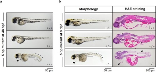
(a) Phenotypes of a 5-bp homozygous deletion (−/−) embryo, heterozygous deletion (+/−) and wild-type (+/+) embryos at 48 hpf are presented. Homozygous mutant embryos displayed severe phenotypes, including smaller eyes, heart and yolk sac edema, smaller yolk extension, curved tail and depigmentation. (b) At 3 dpf, the altered phenotypes and H&E staining were also performed.

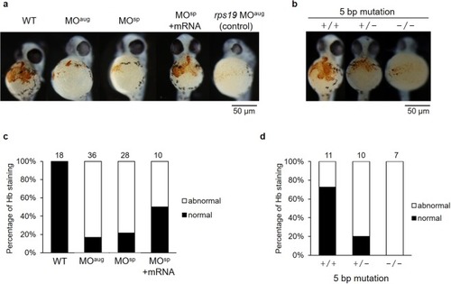
(a) Significant reductions in hemoglobin (Hb) staining in the embryos were observed after rpl10a gene knockdown (MOaug and MOsp), similar to rps19 knockdown, compared to the control; embryos co-injected with rpl10a transcript recovered at 48 hpf. (b) rpl10a embryos mutated using CRISPR-Cas9 knockout showed significant reductions in hemoglobin staining. (+/+: wild-type, +/−: heterozygous, −/−: homozygous mutant). The graph displayed the percentages of normal and abnormal levels of Hb staining in (c) knocked down rpl10a gene and mRNA rescue embryos; (d) 5 bp rpl10a mutant embryos. The dense Hb staining (orange dot) at the wild-type embryo yolk sac was considered normal while a significantly pale orange or slightly colored Hb staining in the yolk sac was evaluated as abnormal. The number of animals quantified in each group are shown on top of the bars.

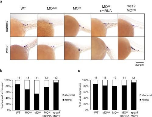
(a) Whole-mount in situ hybridization showing the expression of PGC marker genes, including nanos1 and vasa, after morpholino injection at 25 hpf. The reduction of nanos1 and vasa expression intensity was obviously observed in MOaug-injected embryos. The nanos1 expression was also decreased in MOsp-injected embryos and it was recovered when mRNA was co-injected. The expression levels of nanos1 (b) and vasa (c) were graded as the normal and abnormal percentage. The mean pixel intensity of nanos1 and vasa gene expression of the normal embryos were 24.7 ± 8.7 and 22.6 ± 8.2, respectively. An abnormal level of the gene expression was taken as one with the value less than the mean minus one standard deviation of the gene expression of the wild-type, i.e., less than 16 for nanos1 and 14.4 for vasa gene. The number of animals quantified in each group are shown on top of the bars.

Unillustrated author statements

Acknowledgments
This image is the copyrighted work of the attributed author or publisher, and ZFIN has permission only to display this image to its users. Additional permissions should be obtained from the applicable author or publisher of the image. Full text @ Sci. Rep.