- Title
-
Chronic exposure to ethanol causes steatosis and inflammation in zebrafish liver
- Authors
- Schneider, A.C., Gregório, C., Uribe-Cruz, C., Guizzo, R., Malysz, T., Faccioni-Heuser, M.C., Longo, L., da Silveira, T.R.
- Source
- Full text @ World J Hepatol
|
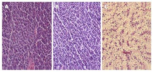
Hematoxylin-eosin staining of liver sections from zebrafish. A: C group (2 wk), the hepatocytes are aligned in cords, absence of fat droplets; B: E group (2 wk), without apparent changes compared with the C group; C: E group (4 wk), enlarged hepatocytes due to fatty infiltration. Magnification: 400 ×. PHENOTYPE:
|
|
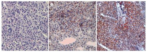
Oil red staining sections of zebrafish liver. A: C group (4 wk), absence of lipid droplets; B: E group (2 wk), mild presence of lipid droplets; C: E group (4 wk), intense lipid accumulation induced by ethanol in hepatocytes. Magnification: 400 ×. PHENOTYPE:
|
|
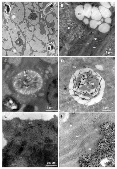
Electron micrographs of liver sections of control (A, C and E) and ethanol exposed groups (B, D and F). A: Polygonal hepatocytes (H), spherical nucleus (n) sinusoid (s), endothelial cell (ec); B: Presence of large amount of glycogen (g) and lipid droplets (ld) in the hepatocytes cytoplasm; C: Intracellular canaliculus with large number of microvilli (a) within; E: It is noted the parallel arrangement of rough endoplasmic reticulum (rer) around the core; D: Myelin figure (mf) inside an intracellular canaliculus; F: Rough endoplasmic reticulum (rer) composed by 8-12 parallel cisterns; H: Hepatocytes; ec: Endothelial cell. PHENOTYPE:
|

ZFIN is incorporating published figure images and captions as part of an ongoing project. Figures from some publications have not yet been curated, or are not available for display because of copyright restrictions. |

ZFIN is incorporating published figure images and captions as part of an ongoing project. Figures from some publications have not yet been curated, or are not available for display because of copyright restrictions. |


