- Title
-
Novel hypophysiotropic AgRP2 neurons and pineal cells revealed by BAC transgenesis in zebrafish
- Authors
- Shainer, I., Buchshtab, A., Hawkins, T.A., Wilson, S.W., Cone, R.D., Gothilf, Y.
- Source
- Full text @ Sci. Rep.
|
AgRP1 and AgRP2 BAC transgenic lines reflect endogenous agrp1 and agrp2 expression patterns. Endogenous mRNA expression of agrp1 and agrp2 was compared to the transgene expression in agrp1:mCherry and agrp2:mCherry larvae, respectively, at 6 dpf. (a,b) ISH analysis for agrp1 mRNA expression in a wild-type larva at 6 dpf. (a) Ventral and (b) lateral views of larvae brains. agrp1 mRNA expression is localized to the ventral periventricular hypothalamus. (c) Lateral view of a 6-dpf agrp1:mCherry transgenic larva. Specific mCherry signal is observed in the ventral periventricular hypothalamus (arrow), which replicates the expression pattern of agrp1 mRNA. (d,e) ISH analysis of agrp2 mRNA expression in a 6-dpf wild-type larva. (d) Dorsal and (e) lateral views of larvae brains. Strong expression of agrp2 mRNA is observed in the pineal gland (top arrow); weaker bilateral agrp2 mRNA expression is observed in the preoptic area (bottom arrow). (f) Lateral view of the brain of a 6-dpf agrp2:mCherry transgenic larva. The expression pattern of mCherry replicates both pineal (top arrow) and preoptic (bottom arrow) agrp2 mRNA expression. (a,d) Anterior to top; (b,c,e,f) anterior to left. H, hypothalamus; HB, hindbrain; OB, olfactory bulb; P, pineal gland; POA, preoptic area; TeO, optic tectum. Scale bar, 100 μm. EXPRESSION / LABELING:
|
|
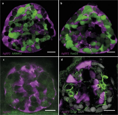
AgRP2 is expressed in uncharacterized pineal cells. (a) Pineal gland of a 5-dpf agrp2:mCherry, Tg(aanat2:EGFP)y9 larva. AgRP2 cells (magenta) do not co-localize with pineal photoreceptor cells (green). (b) Pineal gland of a 5-dpf agrp2:mCherry, Tg(foxd3:EGFP)zf104 larva. AgRP2 cells (magenta) do not co-localize with FoxD3 pineal neurons (green). (c) Double immunostaining of a 5-dpf agrp2:mCherry larva with an antibody against HuC (a neuronal marker) and with anti-RFP. AgRP2 cells (magenta) do not co-localize with HuC-positive cells (green). (d) Immunostaining of a 5-dpf agrp2:EGFP larva with an antibody against GFAP (a marker for pineal interstitial cells) and with anti-EGFP. AgRP2 cells (magenta) do not co-localize with pineal interstitial cells (green). Scale bar, 10 μm. EXPRESSION / LABELING:
|
|
AgRP2 preoptic neurons project towards the pituitary and terminate at the pituitary vasculature. (a) A cross between agrp2:mCherry and Tg(oxt:EGFP)wz01 shows that AgRP2 neurons (magenta) are localized to the preoptic area, adjacent to oxytocin neurons (green), dorsal view of a 5-dpf larva head. (b) Higher-gain imaging of a 5-dpf agrp2:mCherry larva reveals projections of the AgRP2 preoptic neurons towards the pituitary (arrows). (c) Pituitary of a 12-dpf agrp2:mCherry, Tg(pomc:EGFP)zf44 larva, showing AgRP2 projections (magenta) terminating at the adenohypophysis, in close proximity to anterior POMC neurons (green). (d) Pituitary of a 12-dpf agrp2:mCherry, Tg(oxt:EGFP)wz01 larva, showing that projections from AgRP2 (magenta) and oxytocin (green) neurons share the same axonal tracts (arrows) but terminate at different locations within the pituitary: AgRP2 axons terminate at an anterior region whereas oxytocin axons terminate at a posterior region. (e) Pituitary of a 7-dpf agrp2:mCherry, Tg(UAS:SYP-EGFP)biu5 larva expressing synaptophysin (SYP)-EGFP fusion protein and mCherry in AgRP2 neurons. SYP-EGFP protein aggregates in pre-synaptic vesicles at the adenohypophysial AgRP2 terminals. (f) Z-stack projection of a 12-dpf agrp2:mCherry, Tg(kdrl:EGFP)s843 larval pituitary, showing that AgRP2 preoptic projections (magenta) form an interface with the pituitary vasculature (green). (g−g”) show higher magnification of (f). (g) A single focal plane of the pituitary vasculature. (g’) AgRP2 terminals form an arc along the pituitary artery. (g”) Merged image of (g) and (g’). The observed overlap between AgRP2 terminals and the pituitary artery demonstrates a neurovascular interface of AgRP2 terminals with the pituitary vasculature. Anterior to top. AH, adenohypophysis; NH, neurohypophysis; Pit, pituitary; POA, preoptic area. Scale bar, 20 μm. |
|
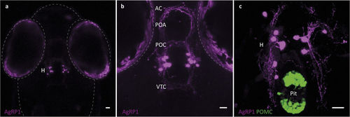
Anatomical organization of AgRP1 neurons. (a) Dorsal view of a 5-dpf agrp1:mCherry larva, showing AgRP1 somata localized at the ventral periventricular hypothalamus (H). (b) Higher magnification of the hypothalamus reveals AgRP1 projections towards the rostral, intermediate and dorsal hypothalamus, the preoptic area (POA), the anterior commissure (AC), the post-optic commissure (POC) and the ventral tegmental commissure (VTC). (c) Ventral view of a 8-dpf agrp1:mCherry; Tg(pomc:EGFP)zf44 larva shows that the AgRP1 neurons (magenta) do not project to the pituitary (Pit, green). Scale bar, 25 μm. |
|
AgRP1 and AgRP2 projections and their interactions. Analysis of potential interactions between AgRP1 and AgRP2 neurons by crossing of agrp1:mCherry with agrp2:EGFP fish. (a) AgRP2 projections (green) towards the adenohypophysis pass alongside AgRP1 somata (magenta). (b−b'') AgRP2 projections potentially form ‘en passant’ synapses with AgRP1 somata. Arrow points to a swelling along the AgRP2 axon in (b'). (b'') Merged image of (b) and (b'), showing co-localization of AgRP2 axonal swelling (green) and AgRP1 soma (magenta). (c) In the pre-optic area, AgRP1 axons (magenta) pass close to AgRP2 somata (green) and share the same axonal tracks with AgRP2 axons. (d,e) Schematic diagrams showing lateral (d) and dorsal (e) views of the anatomical organization of AgRP1 and AgRP2 neuronal systems in 5-dpf larvae. AC, anterior commissure; H, hypothalamus; OB, olfactory bulb; OC, optic chiasm; P, pineal; Pit, pituitary; POA, pre-optic area; POC, post-optic commissure; VTC, ventral tegmental commissure. Scale bar, 10 μm. EXPRESSION / LABELING:
|
|
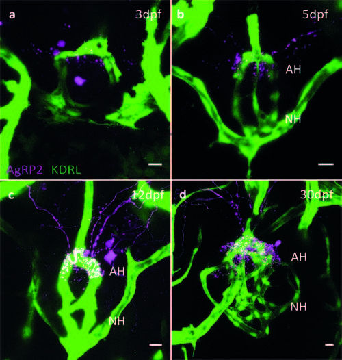
Development of AgRP2 projections (magenta) towards the pituitary vasculature (green). (a) AgRP2 preoptic neuronal projection towards the pituitary vasculature is detected as early as 3 dpf. (b) At 5 dpf, as the pituitary vasculature continues to develop; more AgRP2 projections can be detected. (c) At 12 dpf, a clear interface can be detected between AgRP2 and the pituitary vasculature. (d) At 30 dpf, complexity of the pituitary vasculature increases together with increased number of AgRP2 terminals. EXPRESSION / LABELING:
|

ZFIN is incorporating published figure images and captions as part of an ongoing project. Figures from some publications have not yet been curated, or are not available for display because of copyright restrictions. |


