- Title
-
Cholinergic left-right asymmetry in the habenulo-interpeduncular pathway
- Authors
- Hong, E., Santhakumar, K., Akitake, C.A., Ahn, S.J., Thisse, C., Thisse, B., Wyart, C., Mangin, J.M., and Halpern, M.E.
- Source
- Full text @ Proc. Natl. Acad. Sci. USA
|
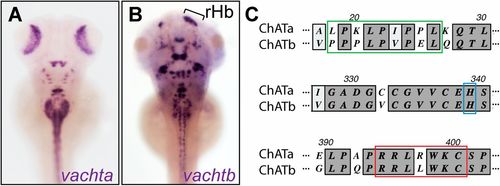
Duplicated zebrafish ChATb. Distribution of vachta (A) and vachtb (B) transcripts in 5-dpf larvae with asymmetrical habenular expression indicated. rHb, right habenula indicated by bracket. (C) Alignment of ChATa and ChATb sequences at the acyltransferase site (green), conserved histidine domain (blue), and putative enzyme catalytical domain (red). EXPRESSION / LABELING:
|
|
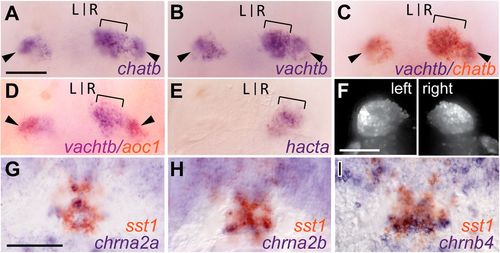
Cholinergic gene expression in zebrafish Hb-IPN; chatb (A) and vachtb (B) transcripts in the right dHb (brackets) and bilaterally in the vHb (arrowheads) of 4-dpf larvae (dorsal views). Double ISH for vachtb with chatb (C) and aoc1 (D). (E) hacta expression only in right dHb. (F) Confocal z-stack images of left and right dHb in Tg(slc17a6b:DsRed) larvae. Transverse sections through sst1-expressing IPN of 4-dpf larvae double-labeled for chrna2a (G), chrna2b (H), and chrnb4 (I). L, left; R, right. (Scale bars: 50 μm.) EXPRESSION / LABELING:
|
|
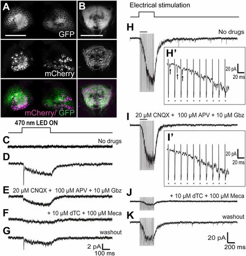
Sustained Hb stimulation induces slow cholinergic current in the vIPN. Confocal images of the dHb (A) and IPN (B) of 5-dpf larvae carrying the TgBAC(gng8:nfsB-CAAX-GFP)c375, TgBAC(gng8:GAL4)c426, and Tg(UAS:ChR2-mCherry)s1985t/+ transgenes. (Scale bars: 50 μm.) Averages of 15 responses evoked in a vIPN neuron by optogenetic stimulation (400 ms) of Hb neurons in control (C) and ChR2-mCherry brains in the absence of drugs (D), presence of 20 μM 6-cyano-7-nitroquinoxaline-2,3-dione (CNQX) + 100 µM amino-5-phosphonopentanoic acid (APV) + 10 μM gabazine (Gbz) (E), with addition of 10 μM (+)-d-tubocurarine chloride (dTc) + 100 μM mecamylamine (Meca) (F), and after washout (G). Examples of responses evoked in a single vIPN neuron by repetitive electrical stimulation of the right Hb (400 ms, 50 Hz) without drugs (H and H2), with 20 μM CNQX + 100 μM APV + 10 μM Gbz (I and I2), with addition of 10 μM dTc + 100 μM Meca (J), and after washout (K). H2 and I2 are higher resolution traces from H and I, respectively, illustrating fast EPSCs (H2, arrows). Asterisks in H2 and I2 mark artifacts of electrical stimulation. EXPRESSION / LABELING:
|
|
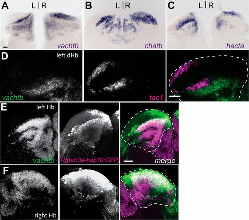
Discrete cholinergic and peptidergic subnuclei of adult dHb. Transverse sections through the Hb of adult brains processed for vactb (A), chatb (B), and hacta (C) expression. (D) Double fluorescent ISH for vachtb and tac1 confirm nonoverlapping cholinergic (green) and tac1-positive (magenta) neuronal populations in the left dHb. Composite z-stack images taken from coronal sections of left (E) and right (F) Hb. Coexpression of vachtb (magenta) and Tg(brn3a-hsp70:GFP) (green). (D–F) White dashed lines delineate dHb, and gray dotted lines delineate vHb. (Scale bars: 50 &mum.) EXPRESSION / LABELING:
|
|
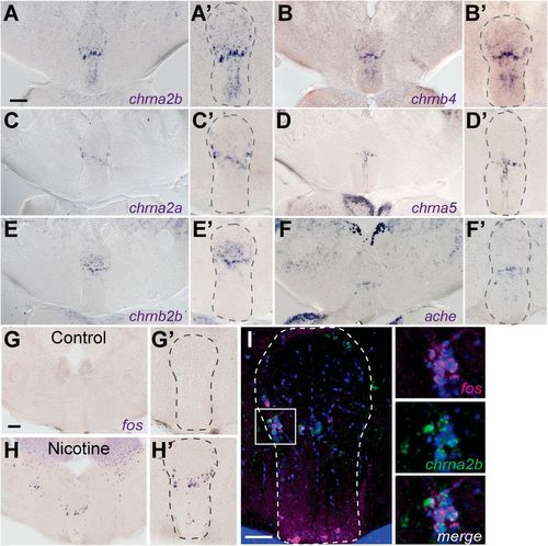
Nicotine-responsive neurons localize to iIPN. Transverse sections of adult brain show chrna2b (A and A2), chrnb4 (B and B2), chrna2a (C and C2), chrna5 (D and D2), chrnb2b (E and E2), and ache (F and F2) transcripts. Control (G and G2) and nicotine-treated (H and H2) fish were processed for fos expression. (Scale bars: A and G, 100 μm.) (A2–H2) Enlarged images of the IPN. (I) Double fluorescent ISH shows fos (magenta) and chrna2b (green) colocalization. DAPI labeling is in blue. The white box in I corresponds to enlarged panels on the right. (Scale bar: 50 μm.) Dotted lines delineate the IPN. EXPRESSION / LABELING:
|
|
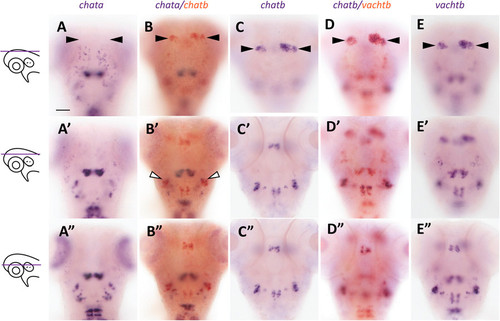
Coexpression of chatb with chata and vachtb. Single and double ISH for chata (A–A22), chata/chatb (B–B22), chatb (C–C22), chatb/vachtb (D–D22), and vachtb (E–E22) in 4-d postfertilization (dpf) larvae, imaged at the designated focal plane (at left). (A–E) Arrowheads mark the Hb. (B2) White arrowheads indicate chata and chatb coexpression in hindbrain neurons. (Scale bar: 50 μm.) EXPRESSION / LABELING:
|

