- Title
-
Excessive activity of cathepsin K is associated with the cartilage defects in a zebrafish model for mucolipidosis II
- Authors
- Petrey, A.C., Flanagan-Steet, H., Johnson, S., Fan, X., De la Rosa, M., Haskins, M.E., Nairn, A.V., Moremen, K.W., and Steet, R.
- Source
- Full text @ Dis. Model. Mech.
|
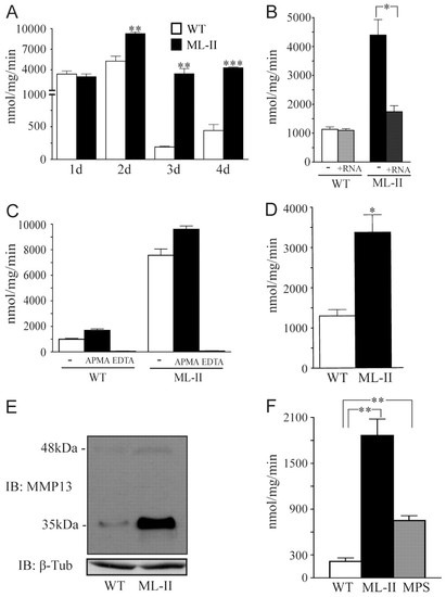
MMP activity is increased in ML-II embryos. (A–C) General MMP activity was measured in WT and ML-II embryo lysates across a developmental time course (1–4 dpf; A), following introduction of GNPTAB mRNA (3 dpf; B) and after incubation with EDTA (inhibits MMPs) or APMA (activates MMPs) for 2 hours (3 dpf; C). (D) Activity of MMP-12 and/or -13 in 3-dpf WT and ML-II embryos measured using specific fluorogenic substrate. (E) MMP-13 immunoblots show increased steady-state levels of both the MMP-13 precursor (48 kDa) and the mature active (35 kDa) forms in 3-dpf ML-II embryo lysates. β-tubulin was used as a loading control. (F) General MMP activity assays in isolated feline synovial fibroblast-like cell lysates. *P<0.05, **P<0.01, ***P<0.001, Student’s t-test. Activity measurements for all conditions were performed on at least three independent embryo lysates. |
|
Cathepsin expression is enriched in the craniofacial skeleton of zebrafish embryos. (A–C) Analysis of enzyme activities in lysates of 3-dpf WT and ML-II embryos separated into heads and tails, as diagrammed in A (dashed line represents cut site) demonstrates that cathepsin activities are primarily increased in the head in ML-II embryos compared with WT (B) rather than in the tail (C); n=3. (D–G) In situ hybridization for cathepsin K expression shows mRNA enrichment in ventral tissues that generally correspond to the pharyngeal skeleton at 2 dpf (D,E) and 3 dpf (F,G) in WT and ML-II embryos. Arrows denote differences in the pattern of expression between WT and ML-II embryos. CB, ceratobranchials; M, Meckels cartilage. (H–J) Immunohistochemical stains for cathepsin K (red) on sections of 3 dpf (H) and 4 dpf (I,J) WT fli1a:EGFP embryos show that it is expressed in chondrocytes and peri-chondrial fibroblasts of the trabecular (H,J) and Meckels cartilages (I). |
|
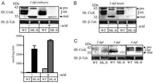
The mature form of cathepsin K is present early and persists in ML-II embryos. (A) Western blot analysis and activity assays of whole embryo lysates with and without acid treatment. (B) Western blot analysis of 3-dpf zebrafish heads with and without acid treatment. (C) Western blot analysis of whole embryo lysates over a developmental time course spanning 2–4 dpf. Procathepsin K (pro; arrowhead), intermediate forms (int; bracket), mature cathepsin K (mat; arrow). |
|
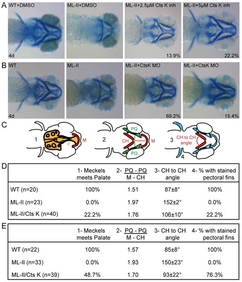
Inhibition of cathepsin K expression or activity results in correction of the ML-II cartilage morphogenesis defects. (A,B) Alcian blue stains of embryos at 4 dpf show that inhibition of either (A) cathepsin K activity using pharmacological agents or (B) expression by MO injection results in significant correction of multiple aspects of the craniofacial defects present in ML-II embryos. Percent values listed represent the number of embryos with these phenotypes. For the drug treatments (A), n=160 embryos in four experiments; for MO experiments (B), n=100 embryos in three experiments. (C) The degree of correction was quantified as follows: (1) whether Meckels (M) cartilage meets the palate (P), (2) the ‘shape’ of the jaw using the ratio of the distance between the palatoquadrate (PQ) bones over the distance from Meckels (M) cartilage to the ceratohyal (CH) bones, (3) the angle between the left and right ceratohyals (CH), and (4) whether the pectoral fins stained positively with Alcian blue. (D,E) The quantitation of the degree of cartilage correction following pharmacological inhibition of cathepsin K activity (D) and SB MO-inhibition of cathepsin K expression (E) are presented. PHENOTYPE:
|
|
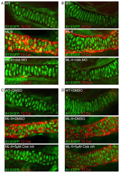
Inhibition of cathepsin K activity reduces type II collagen accumulation in ML-II morphant cartilages. (A–D) Immunohistochemical analysis of type II collagen in the (A,C) trabecular and (B,D) Meckels cartilages from (A,B) WT, ML-II, and ML-II/ctsk MO-inhibited embryos, (C,D) WT, ML-II, and ML-II/cathepsin K inhibitor-treated embryos reveal significant decreases in its expression following inhibition of cathepsin K. PHENOTYPE:
|

Unillustrated author statements |


