- Title
-
Chemical modifier screen identifies HDAC inhibitors as suppressors of PKD models
- Authors
- Cao, Y., Semanchik, N., Lee, S.H., Somlo, S., Barbano, P.E., Coifman, R., and Sun, Z.
- Source
- Full text @ Proc. Natl. Acad. Sci. USA
|
Suppression of pkd2 mutants/morphants by TSA. (A and B) Treatment of 500 nM TSA from the shield stage leads to slight ventral curvature (B) on 2 dpf in wild-type embryos (wt) as compared to embryos treated with DMSO (A). (C–E) TSA can suppress the body curvature of pkd2/hi4166 mutant embryos. (C) shows mutant embryos treated with DMSO from 27 hpf on 2 dpf. Red lines depict the definition of curvature angle. (D) shows embryos treated with 500 nM TSA from 27 hpf on 2 dpf. With this treatment, mutant embryos are indistinguishable from wild-type siblings. (E) Average curvature angle in embryos treated with DMSO, 200 and 500 nM TSA. n = 5, *, P < 0.05, **, P < 0.0005. (F–H) Inhibition of kidney cyst formation in pkd2 morphants (pkd2mo) on 3 dpf by treatment of 100 nm TSA from the bud stage. (F) shows a pkd2 morphants treated with DMSO. The arrow points to the cyst. (F) shows a morphant treated with TSA. Insets show kidney cyst in F or the lack of cyst in G. (H) Average of percentage of embryos with kidney cysts from 3 independent experiments. Twenty-five to fifty embryos were examined in each experiment. ***, P < 0.005. PHENOTYPE:
|
|
Inhibition of Class I HDACs can suppress the phenotypes of pkd2 mutants/morphants. (A and B) Treatment of 20 μm VPA from the shield stage leads to slight ventral curvature (B) on 2 dpf in wild-type embryos (wt) as compared to embryos treated with DMSO (A). (C–E) VPA can suppress the body curvature of pkd2/hi4166 mutant embryos. (C) shows mutant embryos treated with DMSO on 2 dpf. (D) shows mutant embryos treated with 20 μm VPA from 27 hpf on 2 dpf. (E) Average curvature angle in embryos treated with 20 μm VPM and 20 &,mu;m VPA. n = 5, *, P < 0.005, (F–H) Inhibition of kidney cyst formation in pkd2 morphants (pkd2mo) on 3 dpf by treatment of 20 μm VPA from the 20-somite stage. (F) shows a pkd2 morphants treated with 20 μm VPM. The arrow points to the cyst. (G) shows a morphant treated with VPA. White box: area shown in Insets. Insets show kidney cyst in F or the lack of cyst in G. (H) Average of percentage of embryos with kidney cysts from three independent experiments. Twenty-five to one hundred embryos were examined in each experiment. *, P < 0.005. 4166: embryos from pkd2/hi4166 heterozygous cross. PHENOTYPE:
|
|
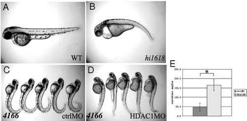
Depletion of hdac1 can suppress the phenotypes of pkd2 mutants. (A and B) Phenotypes of hdac1/hi1618 mutants on 2 dpf in side views. (A) shows a wild-type (WT) sibling, while (B) shows a mutant embryo (hi1618). (C–E) Injection of hdac1 morpholino can suppress body curvature of pkd2/hi4166 mutants on 2 pdf. (C) shows mutant embryos injected with the five base-pair mismatch control oligo. (D) shows mutant embryos injected with the hdac1 morpholino. (E) shows average curvature angle in embryos injected with control or hdac1 morpholino. n = 5, *, P < 0.05. (F–H) Injection of hdac1 morpholino can suppress kidney cyst formation in pkd2 morphants (pkd2mo) on 3 pdf. 4166: embryos from pkd2/hi4166 heterozygous cross; ctrolMO: embryos injected with control morpholino. HDAC1 MO: embryos injected with hdac1 morpholino. PHENOTYPE:
|
|
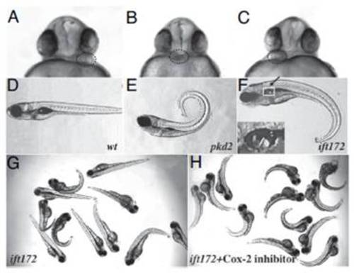
A chemical modifier screen for the body curvature and LR asymmetry phenotypes of zebrafish PKD models. (A–C) Embryos at 30 hpf with heart (red circles) at the Left side (A), Middle (B) and Right side (C) shown in ventral views. (D–F) Embryos with different body curvature phenotype on 5 dpf. (D) shows a wild-type embryo (wt) with straight body. (E) shows a pkd2/hi4166 mutant (pkd2) with dorsally curved body axis. (F) shows an ift172/hi211 mutant embryo (ift172) with ventrally curved body axis and kidney cyst (arrow and Inset). White box: area shown in Inset. (G and H) An example of a compound that affects body curvature. Treatment of 20μMCox-2 inhibitor II changes ventral curvature of ift172/hi2211 mutants and wild-type siblings to dorsal curvature. (G) shows embryos on 3 dpf from ift172/hi2211 heterozygous carriers treated with DMSO on 3 dpf. (H) shows embryos treated with the compound. PHENOTYPE:
|
|
Treatment of TSA causes ventral body curvature in a dosage dependent manner. (A) Embryos were treated with DMSO, 100, and 200 nM TSA from the 30% epiboly stage. (B) pkd2/hi4166 mutant embryos on 2 dpf treated with 200 nM TSA from 27 hpf, compared with embryos treated with DMSO (Fig. 1C) and 500 nM TSA (Fig. 1D). |
|
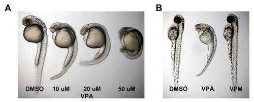
Treatment of VPA causes ventral body curvature in a dosage dependent manner. (A) Embryos were treated with DMSO, 10, 20, and 50 μM VPA from the 30% epiboly stage. (B) Embryos treated with DMSO, 20 μM VPA, or 50 μM VPM from the 30% epiboly stage on 2 dpf. |
|
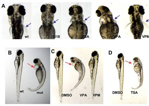
Comparison of phenotypes caused by hdac1 depletion and VPA and TSA treatment. (A) Stunted pectoral fin (blue arrow) development on 3 dpf in a hdac1/hi1618 mutant embryo (1618), a wild-type embryo treated with 300 nM TSA (TSA), 20 μM VPA (VPA), and 50 μm (VPM) from the 30% epiboly stage. (B) Pericardiac edema (red arrow) on 3 dpf in a hdac1/hi1618 mutant embryo. WT: wild-type sibling. mut: hdac1/hi1618 mutant. (C) Treatment of 300 nM TSA from the 30% epiboly stage leads to pericardiac edema (red arrow) on 3 dpf, while similar treatment with 50 μM VPM does not. (D) Treatment of 20 μM TSA from the 30% epiboly stage leads to pericardiac edema (red arrow) on 3 dpf. |
|
Automated classification of the body curvature phenotype. The original image (A) is first passed trough a classification algorithms trained for separating the fish-pixels from the background. The output (B) is then denoised with a convolutional filter and the result (C) is passed to the detection stage. Fish classified as straight by the program is marked with a red line, while curved is labeled with a blue line (D). A misclassification caused by two fish lumping together can be seen in D (arrow). |


