Fig. 5
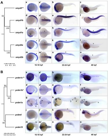
Fig. 5 Somite/muscle-expressed SET domain genes and their evolutionary relationships. The phylogenetic relationships of the genes were indicated with the trees constructed based on the SET domains of the encoded proteins and rooted with zebrafish Smyd4 and Prdm14 proteins as outgroups, respectively. Lateral views (anterior to the left) of embryos at 16–18 hpf (a, d, g, j and m), 22–24 hpf (b, e, h, k and n) and 48 hpf (c, f, i, l and o) are presented. (a', d', g', j' and m') Dorsal views of the embryos in a, d, g, j and m. (A) Zebrafish smyd1a, smyd1b, smyd2a and smyd2b genes show somite/muscle-specific expression patterns and form a close paralog group with the smyd3 gene (double asterisks), which shows a ubiquitous expression pattern (a–c). Note the relatively low expressions of smyd1a at early stage (18 hpf; g) and smyd2a and smyd2b at late stage (48 hpf; l and o). (B) Expression patterns of the second paralog group. prdm1a is specifically expressed in anterior somites and adaxial cells at 18 hpf (g and g') and 24 hpf (h). Besides, it is also expressed in hatching gland (g), branchial arch, fin fold (g, g' and h), fin buds, cloaca (h and i) and retina (i). prdm1b (asterisk) is highly expressed in somites at 24 hpf (e) and in retina at 48 hpf (f). prdm1c (double asterisks) is ubiquitously expressed (a–c). prdm4 is highly expressed in somites and retina (k and l). prdm15 is expressed in muscle pioneer cells (m, m' and n). ac, adaxial cells; ba, branchial arch; cl, cloaca; fb, fin buds; ff, fin fold; hg, hatching gland; mp, muscle pioneer; re, retina; s, somite.

