Fig. 7
- ID
- ZDB-FIG-240716-19
- Publication
- Lu et al., 2024 - Localisation and function of key axonemal microtubule inner proteins and dynein docking complex members reveal extensive diversity among vertebrate motile cilia
- Other Figures
- All Figure Page
- Back to All Figure Page
|
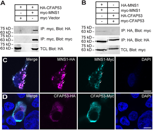
Biochemical interaction between MNS1 and CFAP53. (A) Myc-tagged MNS1 immunoprecipitated with HA-tagged CFAP53. (B) MNS1, but not CFAP53, exhibited self-interaction. (C) MNS1-Myc and MNS1-HA formed filament-like structures in HEK293T cells (arrows). MNS1-HA was stained with anti-HA antibody (magenta); MNS1-Myc was stained with anti-Myc antibody (cyan). (D) CFAP53-Myc and CFAP53-HA showed diffuse staining in HEK293T cells. CFAP53-HA was stained with anti-HA antibody (magenta); CFAP53-Myc was stained with anti-Myc antibody (Cyan). Scale bars: 5 µm. Immunoprecipitation was verified with two biological replicates. Immunofluoresence data represent two technical replicates with 100 cells analysed per replicate. |

