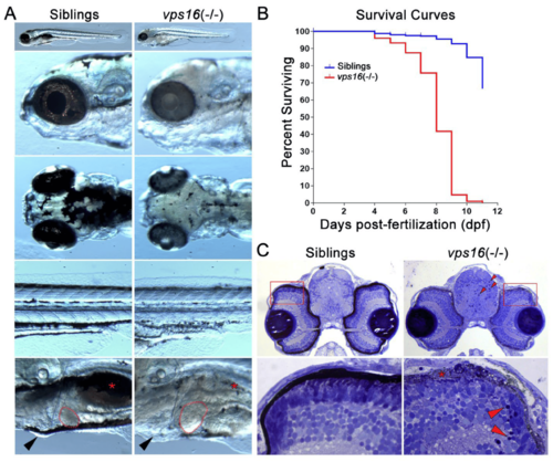
vps16(-/-) mutants can be phenotypically distinguished from wild-type siblings. (A) Images of wild-type sibling (left) and vps16(-/-) (right) larvae displaying distinctive pigmentation patterns at 7 dpf. Sibling larva (left) with characteristic darkly pigmented melanophore in RPE layer within eye and darkly pigmented melanophore aggregates on top of head and near yolk sac extension on side of body. Location of normally pigmented swim bladder (red asterisk), size of liver (outlined in red), and absence of edema (black arrowhead) shown in sibling larvae. vps16(-/-) mutant larva (right) showing severe hypopigmentation in RPE and reduced pigmentation on head and near yolk sac extension. vps16(-/-) mutant displaying hypopigmented and reduced swim bladder (red asterisk), hepatomegaly (outlined in red), and pericardial edema (black arrowhead). (B) Survival curve of siblings (blue) and vps16(-/-) mutants (red) depicting death of all mutants by 11 dpf. (C) Histological sections of 7 dpf sibling and vps16(-/-) larval forebrain near optic nerve showing pyknotic nuclei in mutant brain and retina (red arrowheads). vps16(-/-) larval retina shows degeneration and absence of pigmented RPE and truncated photoreceptors (asterisks) and lack of distinct organization of photoreceptor outer segments.
|
