FIGURE

Figure 6

ID
ZDB-FIG-230625-20
Publication
Amin et al., 2023 - Short-Term TERT Inhibition Impairs Cellular Proliferation via a Telomere Length-Independent Mechanism and Can Be Exploited as a Potential Anticancer Approach
Other Figures
All Figure Page
Back to All Figure Page
Figure 6

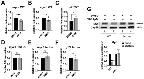
BIBR treatment downregulated the expression of zebrafish myca and mycb and upregulated p21. Twelve hours post fertilization (hpf), zebrafish wild-type (WT) and tert mutant (terthu3430/hu3430; tert −/−) embryos were treated with 2 μM BIBR or DMSO as a control for 12 h, from 12 to 24 hpf. Levels of relative mRNA expression for the indicated genes in WT (AC) and tert mutant (DF) embryos are shown. Data represent the mean and SD (bar) from three separate experiments. (G) Representative Western blots showing total protein levels of Myc in WT and tert mutant zebrafish embryos upon treatment with 2 μM BIBR for 12 h. Glyceraldehyde-3-phosphate dehydrogenase (Gapdh) was used as a loading control. The original Western blots are shown in File S1. The graph shows the values in arbitrary units of densitometric analysis performed with ImageJ software. Data represent the mean and SD (bar) from two separate experiments. A significant difference between values in BIBR-treated embryos vs. DMSO-treated embryos is shown: * p < 0.05; *** p < 0.001; ns: not significant.

Expression Data

Expression Detail
Antibody Labeling
Phenotype Data

Phenotype Detail
Acknowledgments
This image is the copyrighted work of the attributed author or publisher, and ZFIN has permission only to display this image to its users. Additional permissions should be obtained from the applicable author or publisher of the image. Full text @ Cancers