
Fig. 2.
- ID
- ZDB-FIG-230420-156
- Publication
- Peterman et al., 2023 - Zebrafish cutaneous injury models reveal Langerhans cells engulf axonal debris in adult epidermis
- Other Figures
- All Figure Page
- Back to All Figure Page
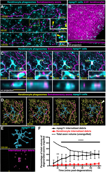
mpeg1+ cells, not keratinocytes, engulf the majority of axon debris following axon degeneration. (A) Confocal images from a time-lapse of scale pluck-induced axon degeneration from an adult expressing reporters for keratinocyte phagosomes [Tg(krt4:EGFP-2xFYVE);TgBAC(ΔNp63:EGFP-2xFYVE)] and somatosensory axons [Tg(p2rx3a:mCherry)]. Arrowheads show examples of colocalization. Time denotes mm:ss relative to the onset of axon degeneration. See Movie 2. (B) Lateral confocal image (top) and reconstructed cross-section (bottom), showing that mpeg1+ cells (cyan) densely populate the scale epidermis and reside beneath the krt4+ layer (magenta). (C) Confocal images from a time-lapse of axon degeneration from an adult expressing reporters for keratinocyte phagosomes [Tg(krt4:EGFP-2xFYVE);TgBAC(ΔNp63:EGFP-2xFYVE)], somatosensory axons [Tg(p2rx3a:mCherry)] and mpeg1+ cells [Tg(mpeg1:NTR-EYFP)] before and during scale pluck-induced axon degeneration. Yellow dotted lines in insets denote the plane reconstructed in the xz projections. Time denotes mm:ss relative to the onset of axon degeneration. See Movies 3 and 4. (D) Surface views from Imaris of the panels in C showing keratinocyte phagosomes (yellow), mpeg1+ cell (cyan) and somatosensory axons (magenta). Arrowhead indicates intact axon. (E) Surface view used in Imaris from C (120:00) for volume-engulfed quantifications. (F) Quantification of total axon volume engulfed over time by keratinocytes and mpeg1+ cells. Two-way ANOVA followed by Bonferroni tests determined significance of differences between mpeg1+ cells and keratinocytes. **P<0.01, ****P<0.0001. n=10-16 cells/ROIs from n=12 scales from N=9 fish. Scale bars: 5 μm [A, insets, C, C (xz projection), E], 10 μm (A,D), 30 μm (B), 2 μm (C, insets). Error bars in F represent s.d. |