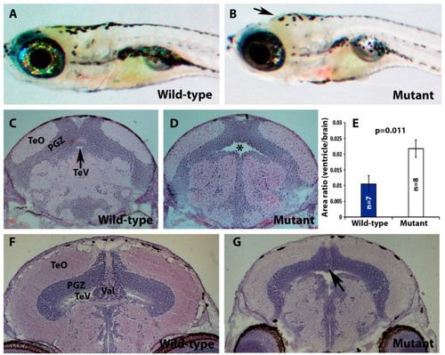
pomt2 mutant zebrafish exhibit hydrocephalus, hypoplasia of the cerebellum, and muscular dystrophy. Sections of the brain and skeletal muscle were stained with H&E. (A,B) Lateral view of a wild-type and a pomt2 mutant zebrafish both dead 3 weeks post fertilization. Note the domed head in the mutant (arrow). (C,D) H&E staining of the rostral region of the optic tectum in zebrafish at 2-mpf. Note pomt2 mutant zebrafish showing an enlarged tectal ventricle (asterisk in (D)). (E) Ratio of ventricular area to total brain cross-sectional area. Note the increased ratio in the mutant. (F,G) H&E staining of the caudal region of the optic tectum in zebrafish at 2-mpf. Homozygous pomt2sny5+13 mutant exhibited hypoplasia of the cerebellum (arrow). (H,I) H&E of skeletal muscle at 2-mpf. Note presence of dystrophic myofibers including necrotic fibers (asterisks), centrally located nuclei (arrowhead), and variably sized fibers. Scale bar in I: 50 µm for (C,D), 60 µm for (F,G), 28 µm for (H,I). Abbreviations: TeO = tectum opticum; PGZ = periventricular gray zone; TeV = tectal ventricle; Val = valvular cerebellum.
|
