
Fig. 1
- ID
- ZDB-FIG-220815-1
- Publication
- Ethiraj et al., 2022 - Colorimetric and fluorescent TRAP assays for visualising and quantifying fish osteoclast activity
- Other Figures
- All Figure Page
- Back to All Figure Page
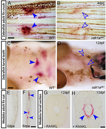
Identification of activated osteoclasts using conventional TRAP stain. A-H) Brightfield images of zebrafish (A-D) and medaka (E-H) adult fins (A-B,E,F), or 12dpf larvae (C,D,G H) processed by colorimetric TRAP staining. Individuals are either wild-type (WT; A, C, E, F), homozygous csf1raj4e1 mutants (B,D) or transgenic for rankl:HS:cfp (G,H). Images are of whole-mount fins at 4 dpc (A,B), whole-mount ventral views (C, D) or longitudinal (E) or transverse cryosections (G,H). Fins in (E,F) have been amputated and stained immediately (0dpa; E) or after 6 days (dpa; F). The larva in (H) has been subjected to heat shock to express RANKL, while the larva in (G) was not heat shocked. Red precipitate (blue arrowheads) indicates TRAP locations at fractures (A), pharyngeal teeth of the fifth ceratobranchial bone (C), in regenerating fin rays at 6 days post amputation (6dpa; F) or around notochord in larvae with excess RANKL induced by heat shock (H). TRAP staining is absent when there no RANKL overexpression (G), at the start of fin regeneration (0dpa; E) or following genetic ablation of osteoclasts (B, D; open blue arrowheads), indicating specificity. Scale bars: A) 200 μm; C) 100 μm; F,H) 50 μm. |