Fig. 3
|
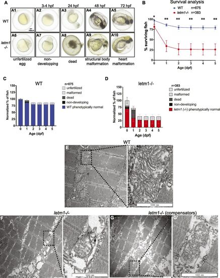
letm1 KO impacts embryonic development, survival and mitochondrial morphology. (A) Images of WT and letm1−/− zebrafish embryos. Lower panel display representative images of the different categories used in the phenotypic analysis. (A1–A5) Representative images of wild-type, phenotypically normal fish. (A6–A10) Representative images of letm1−/−, (A6) unfertilized egg, (A7) non-developing, (A8) dead, (A9) structural body malformation, and (A10) heart malformation. Scale bar: 100 μm. (B) Survival analysis of zebrafish wild-type (WT) and letm1−/− from 0 to 5 day post fertilization (dpf). Quantification of the number of surviving fish per day shown in percentage. The total number of fish analyzed was nWT = 675 and nletm1−/− = 363. Graph shows mean ± SEM. Welch t test, *P < 0.05. (C, D) Phenotypic analysis of zebrafish WT (C) and letm1−/− (D) from 0 to 5 dpf. (E, F, G) Transmission electron microscopy images of 4 dpf zebrafish larvae: WT (E) Longitudinal section of muscle fibers show dense mitochondrial; malformed letm1−/− (F) have less cristae in intramyofibrillar mitochondria, arrow points to detached outer membrane, asterisk to a vacuole-like shape, possibly indicating the transition between damaged, and degraded mitochondria; letm1−/− compensators, with denser mitochondria than in non-compensator letm1−/− and apparently not fusing with vacuoles (G). Scale bar 1,000 nm. (E’, F’, G’) Magnification of the boxed dotted line area. |
| Fish: | |
|---|---|
| Observed In: | |
| Stage Range: | 1-cell to Day 5 |

