
Fig. 6
- ID
- ZDB-FIG-220620-28
- Publication
- Cairelli et al., 2022 - Fluid mechanics of the zebrafish embryonic heart trabeculation
- Other Figures
- All Figure Page
- Back to All Figure Page
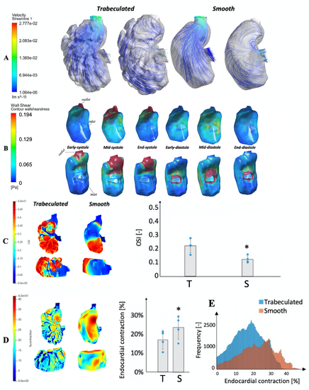
Fig 6. Comparison of hemodynamic parameters between traeculated and smooth embryonic ventricles. (A) Velocity streamlines for both trabeculated and smoothed wall simulations of a 3dpf zebrafish embryonic heart from lateral views, at (left) the end-diastolic phase and (right) the mid-systolic phase. (B) Contour maps of endocardial WSS in the same embryonic ventricle of Fig 2, but with a totally smooth geometry, over the cardiac cycle with the assumption that fluid has the viscosity of plasma (1.5cP). Top row: ventral view of the outer curvature of the ventricle; bottom row: dorsal view of the inner curvature (C) Spatial pattern and surface-averaged magnitudes of oscillatory shear index (OSI) for both trabeculated (T) and smoothed (S) wall simulations of a 3dpf zebrafish embryonic heart, from lateral and ventral views. (D) Spatial pattern and surface averaged-magnitudes of endocardial contractile surface area strains (end-diastole to end-systole), for both the trabeculated (T) and smoothed (S) simulations, from the same views. (E) Histograms of the endocardial contraction across surface locations for the trabeculated and smooth models of the same ventricular chamber.* p values were found to be at the minimum possible with the small sample size (n = 4, p = 0.0625). |