
Fig. 1
- ID
- ZDB-FIG-220620-23
- Publication
- Cairelli et al., 2022 - Fluid mechanics of the zebrafish embryonic heart trabeculation
- Other Figures
- All Figure Page
- Back to All Figure Page
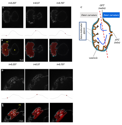
Fig 1. Microscope images and anatomic 3D models of the developing zebrafish endocardium. (A,D) Raw microscope images of (A) the whole ventricle, and (D) a single intra-trabecular space at 25%, 50% and 75% of the cardiac cycle. (B, E) Segmentation of (B) the ventricle and (E) a single intra-trabecular space, superimposed on the raw images, at the same time points as (A) and (D) (also shown in S1 Movie). The 3D reconstructed volumes are in red, while the regions of the 3D reconstruction close to the plane of the shown image are plotted in cyan on single 2D slice extracted from 4D image stacks of a zebrafish embryo from the Tg(fli1a:Gal4ff;UAS:EGFP-CAAX) line at 3 dpf. (C) Schematization of the intra-trabecular geometry, adapted from [15]. A, atrium; V, ventricle; ITS, intra-trabecular space. Here, t denotes time, while T denotes cardiac cycle duration. |