
Fig. 8
- ID
- ZDB-FIG-210602-23
- Publication
- Frétaud et al., 2020 - A new reporter zebrafish line unveils heterogeneity among lymphatic endothelial cells during development
- Other Figures
- All Figure Page
- Back to All Figure Page
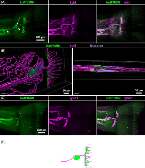
Tail fin lymphatics are batf3MIN positive A, Maximum projection of tail in double Tg(batf3MIN:eGFP) x Tg(kdrl:Hsa.HRAS-mCherry) transgenic fish at 21 dpf. B, Lateral (left panel) and dorsal (right panel) view of the 3D rendered tail showing the lymphatic heart (green, arrow) associated to autofluorescent muscle fibers (blue) and blood vasculature (magenta). Caudal fin lymphatic vessels were surrounding blood vessels of the caudal fin extending towards the rays. C, Maximum projection of tail in double Tg(batf3MIN:eGFP) x Tg(lyve1:dsRed2nz101) transgenic fish at 21 dpf. D, Schematic diagram of caudal fin lymphatics and blood vasculature in lateral view at 21 dpf |