
Fig. 2
- ID
- ZDB-FIG-210602-17
- Publication
- Frétaud et al., 2020 - A new reporter zebrafish line unveils heterogeneity among lymphatic endothelial cells during development
- Other Figures
- All Figure Page
- Back to All Figure Page
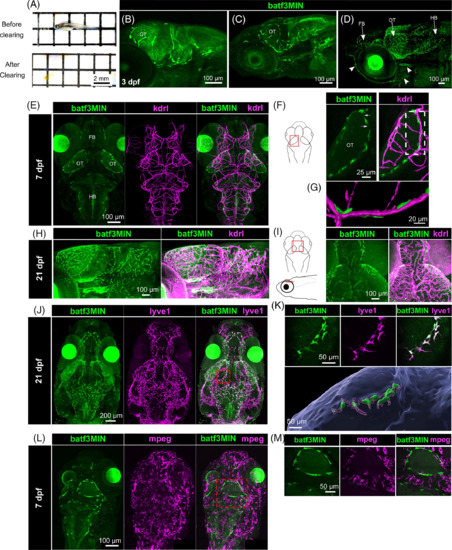
Cranial and facial lymphatics endothelial cells express eGFP in the Tg(batf3MIN:eGFP) reporter line. A, Fixed 21 dpf zebrafish (upper panel) was depigmented and cleared to obtain totally transparent fish (bottom panel). B-D, Two-photon lateral images of head from 3, 7, and 21 dpf Tg(batf3MIN:eGFP) zebrafish. Arrowheads indicate facial lymphatics. E, Dorsal images of head from double Tg(batf3MIN:eGFP) x Tg(kdrl:Hsa.HRAS-mCherry) transgenic fish at 7 dpf, F, Schematic diagram of zebrafish head in dorsal view. Red box indicated the region of optic tectum acquired. Maximum projection of the left optic tectum in double Tg(batf3MIN:eGFP) x Tg(kdrl:Hsa.HRAS-mCherry) transgenic fish at 7 dpf. Arrows show thin projections linking GFP expressing cells to each other G, 3D reconstruction of dashed line highlighted region shown on the F right panel. H, Lateral maximum projection of the brain in double Tg(batf3MIN:eGFP) x Tg(kdrl:Hsa.HRAS-mCherry) transgenic fish at 21 dpf. I, Schematic diagram of zebrafish brain in dorsal and lateral view. Red box indicated the region imaged. Maximum projections of a brain region acquired in double Tg(batf3MIN:eGFP) x Tg(kdrl:Hsa.HRAS-mCherry) transgenic fish at 21 dpf. J, Dorsal images of head from Tg(batf3MIN:eGFP) x Tg(lyve1:dsRed2nz101) transgenic fish at 21 dpf. Red box indicated the region of the optic tectum presented in k. K, Upper panel: maximum projection of a region of the optic tectum in 21 dpf double Tg(batf3MIN:eGFP) x Tg(lyve1:dsRed2nz101) transgenic fish. Lower panel: 3D reconstruction region presented in upper panels (batf3MIN), magenta (lyve1) and blue (brain surface). L, Dorsal images of head from Tg(batf3MIN:eGFP) x Tg(mpeg:mCherryF) transgenic fish at 7 dpf. Red box indicated the region of the brain presented in m. M, Dorsal view of a region of the brain in a 7 dpf Tg(batf3MIN:eGFP) x Tg(mpeg:mCherryF) transgenic fish. FB, forebrain; OT, optic tectum; HB, hindbrain |