|
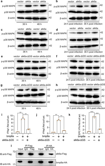
Bmp8a activates the IFN expression through Alk6a.a, c Immunoblot analysis of phosphorylated (p-) p38 MAPK after transfected with 2 μg alk6a or empty vector in ZFL (a) or EPC (c) cells. The cells were collected at 36 or 48 h of post-transfection for Immunoblot analysis. b, d Immunoblot analysis of phosphorylated (p-) p38 MAPK after transfected with 2 μg alk6a or empty vector in ZFL (b) or EPC (d) cells for 24 h, followed by infection with GCRV (5 × 10⁴ TCID50 per ml) for another 24 h or 36 h. e, g Immunoblot analysis of phosphorylated (p-) p38 MAPK after transfected with 2 μg alk6a-ΔGS or empty vector in ZFL (e) or EPC (g) cells. The cells were collected at 36 or 48 h of post-transfection for Immunoblot analysis. f, h Immunoblot analysis of phosphorylated (p-) p38 MAPK after transfected with 2 μg alk6a or empty vector in ZFL (f) or EPC (h) cells for 24 h, followed by infection with GCRV (5 × 10⁴ TCID50 per ml) for another 24 or 36 h. i–k EPC cells were cotransfected with IFN-φ1pro-luc (200 ng, i), IFN-φ3pro-luc (200 ng, j) or EPC IFNpro-luc (200 ng, k), and bmp8a (100 ng) together with or without the dominant negative plasmids alk6a-ΔGS (100 ng). At 48 h of post-transfection, the cells were collected for luciferase assays. Renilla luciferase was used as the internal control. l Co-immunoprecipitation and immunoblot analysis of EPC cells cotransfected with alk6a-Flag (1 μg) and bmp8a-HA (1 μg). Data were from three independent experiments and were analyzed by Student’s t-test (two-tailed) for comparison of two groups or one-way ANOVA followed by Games–Howell posthoc tests for comparison of multiple groups. All data were presented as mean ± SD (**p < 0.01, ***p < 0.001).
|