|
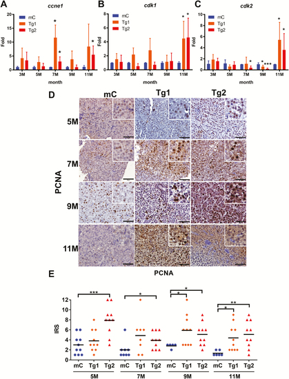
RPIA-transgenic fish increase expression of cell cycle/proliferation markers and PCNA nuclear staining. (A–C) QPCR was used to measure the relative mRNA fold induction of the cell cycle/proliferation markers ccne1, cdk1 and cdk2 in two independent lines of RPIA-transgenic fish (Tg1 and Tg2) compared with the control fish (mC) at 3, 5, 7, 9 and 11 months of age. (D) Representative images of PCNA IHC result for the control (mC) and Tg1 and Tg2 fish aged 5–11 months. All of the magnifications were ×400. Scale bar: 50 μm. (E) Statistical analysis of the PCNA IHC staining results from Tg1 and Tg2 compared with control, as determined by the IRS scoring system. Statistical analysis was performed by two-tailed Student’s t-tests. The symbol ‘*’ represents significance between RPIA-transgenic fish and GFP-mCherry control. P < 0.05 was considered to be statistically significant; *: 0.01 < P ≤ 0.05; **: 0.001 < P ≤ 0.01; ***: P ≤ 0.001.
|