FIGURE

Figure 6

ID
ZDB-FIG-190723-971
Publication
Anbalagan et al., 2019 - Robo2 regulates synaptic oxytocin content by affecting actin dynamics
Other Figures
All Figure Page
Back to All Figure Page
Figure 6

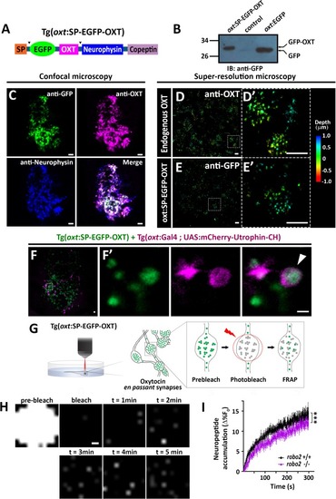
Specificity of antibodies to oxytocin and neurophysin.

(A) Schema of protein coded by oxt gene. Antibodies anti-OXT and PS45 were targeted to processed OXT and neurophysin respectively. SP denotes signal peptide, arrow indicates cleavage site. Validation of the EGFP-OXT fusion protein is shown in Figure 3H. (B–F) Confocal Z-stack images showing Fluorescent in situ hybdridization (FISH) of 3 days post-fertilization (dpf) larvae using probes for avp (B) or oxt (C) mRNA (red), followed by PS45 staining. The neurosecretory preoptic (NPO) area in which oxytocin neurons were labeled is shown. Confocal maximum intensity projection (MIP) images showing immunostaining of 5-dpf old transgenic Tg(oxt:EGFP) larvae using antibodies directed against GFP and neurophysin (NP). The whole neurohypophyseal tract (D) and the hypophyseal projections (E) are shown. Scale: 10 μM.

Expression Data
Gene:
Antibody:
Fish:
Anatomical Term:
Stage: Day 5

Expression Detail
Antibody Labeling
Phenotype Data

Phenotype Detail
Acknowledgments
This image is the copyrighted work of the attributed author or publisher, and ZFIN has permission only to display this image to its users. Additional permissions should be obtained from the applicable author or publisher of the image. Full text @ Elife