
Fig. 2
- ID
- ZDB-FIG-190701-9
- Publication
- Cal et al., 2019 - Countershading in zebrafish results from an Asip1 controlled dorsoventral gradient of pigment cell differentiation
- Other Figures
- All Figure Page
- Back to All Figure Page
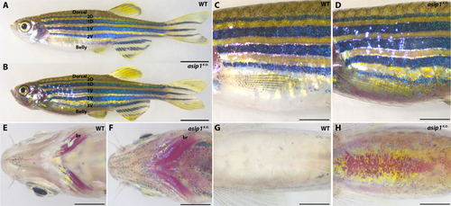
Adult dorso-ventral countershading pattern is disrupted in asip1K.O.. Lateral (A,B), anterior-lateral (C,D), ventral head (E,F) and ventral belly (G,H) views of 180 dpf WT and asip1K.O. zebrafish. (A,B) The pigment pattern of WT zebrafish is a striped pigment pattern with dark stripes and light interstripes. Each dark stripe is named with a code: two primary stripes are called 1D and 1 V, and the two secondary stripes are named 2D and 2 V. The asip1K.O. display an extra 3 V dark stripe. The asip1K.O. phenotype is characterized by a darker belly than WT. (C,D) The striped pigment pattern was almost unaltered in asip1K.O. fish, except that the 2 V stripe is wider than in WT, and the ventral dark stripe 3 V is better developed anteriorly. The darker belly of asip1K.O. compared to WT sibling fish is clearly evident. (E,F) In WT, melanophores are infrequent around the jaws and branchiostegals; however, branchiostegal, jaw and operculum regions are clearly hyperpigmented in asip1K.O.. (G,H) Melanophores are virtually absent in WT belly; thus, WT ventral region shows bright white color as a result of high number of iridophores in the abdominal wall. However, asip1K.O. shows a consistent hyperpigmentation, with many melanophores and xanthophores in the ventral skin; the abdominal wall also seems to be affected, with reduced extent of iridophores and looking much yellower than WT. Scale bar: (A,B) 5 mm, (C–H) 2 mm. Abbreviation: br, branchiostegal.
|
| Fish: | |
|---|---|
| Observed In: | |
| Stage: | Adult |