
Fig. 3
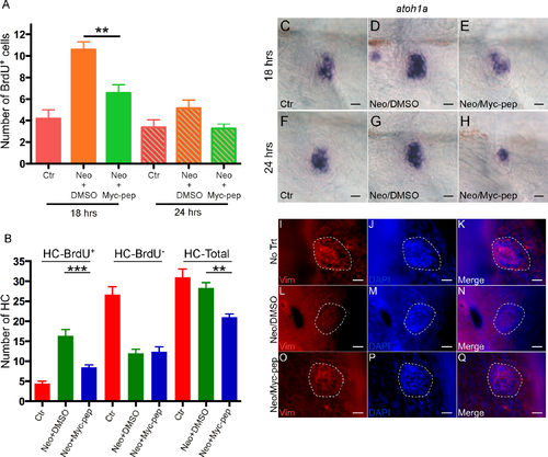
Inhibition of c-Myc blocked proliferation and down-regulated vimentin during HC regeneration. (A) A significant reduction in the number of proliferating neuromast cells (BrdU+) was seen 18 or 24 hrs after c-MYC inhibitor Int-H1-S6A, F8A (Myc-pep, 100 nM) treatment. Ctr, time-matched control fish without neomycin treatment. (B) c-MYC peptide inhibitor mainly blocked proliferation-derived HCs (HC-BrdU+) at 72 hrs. ***p<0.001; **p<0.01. A and B, n = 15 for each group. Two independent experiments were performed with similar results. (C-H) In situ hybridization of atoh1a on 5-dpf zebrafish neuromasts 18 (C-E) or 24 hrs (F-H) after neomycin treatment, with either Myc-pep or DMSO in the media. Ctr, time-matched control fish without neomycin treatment. atoh1a up-regulation by neomycin treatment was blocked in the Myc-pep treatment group (D-E, G-H). (I-Q) Vimentin was down-regulated by c-Myc during HC regeneration. 5-dpf neuromasts were labeled with vimentin and DAPI 18 hrs after neomycin treatment, with either Myc-pep (O-Q) or DMSO (L-N) in the media. No Trt, time-matched control fish without neomycin treatment (I-K). Scale bars: 10 μm. |
| Gene: | |
|---|---|
| Antibody: | |
| Fish: | |
| Condition: | |
| Anatomical Term: | |
| Stage: | Day 5 |
| Fish: | |
|---|---|
| Condition: | |
| Observed In: | |
| Stage: | Day 5 |